Topics
Sexual Reproduction in Flowering Plants
- Sexual Reproduction
- Life Cycle of Angiosperms
- Flower
- Types of Flowers
- Events in Sexual Reproduction > Pre-fertilization Events
- Structure and Development of Anther
- Microsporogenesis
- Structure and Development of Male Gametophyte
- Structure and Development of Ovule
- Types of Ovules (Based on Orientation)
- Types of Ovules (Based on Integuments)
- Megasporogenesis
- Development of Female Gametophyte or Embryo Sac
- Male Vs Female Gametophytes in Angiosperms
- Pollination
- Self-pollination
- Autogamy
- Cross-pollination
- Geitonogamy
- Outbreeding Devices
- Agents and Types of Cross-pollination
- Anemophily
- Hydrophily
- Entomophily
- Ornithophily
- Cheiropteriphily
- Malacophily
- Wind Vs Insect-pollinated flowers
- Self-pollination Vs Cross-pollination
- Events in Sexual Reproduction > Fertilization (Syngamy)
- Artificial Hybridization or Artificial Fertilization
- Pollen Pistil Interaction
- Double Fertilization and Triple Fusion
- Events in Sexual Reproduction > Post-Fertilization Structures and Events
- Endosperm
- Dicotyledonous Embryo
- Monocotyledonous Embryo
- Parthenocarpy
- Polyembryony
- Apomixis
- Formation of Seed and Fruit
- The Fruit
- The Seed
Reproduction
Reproduction in Organisms
- Reproduction
- Modes of Reproduction
- Asexual Reproduction
- Asexual Reproduction in Unicellular Organisms > Binary Fission
- Binary Fission > Simple Binary Fission
- Binary Fission > Transverse Binary Fission
- Binary Fission > Longitudinal Binary Fission
- Asexual Reproduction in Unicellular Organisms > Multiple Fission
- Asexual Reproduction in Unicellular Organisms > Budding
- Asexual Reproduction in Multicellular Organisms > Budding
- Asexual Reproduction in Animals > Gemmule Formation
- Asexual Reproduction in Multicellular Organisms > Regeneration
- Asexual Reproduction in Multicellular Organisms > Fragmentation
- Asexual Reproduction in Multicellular Organisms > Spore Formation
- Binary Vs Multiple Fission
- Vegetative Reproduction or Vegetative Propagation
- Natural Vegetative Propagation
- Artificial Vegetative Propagation
- Sexual Reproduction
- Monocarpic Vs Polycarpic Plants
- Events in Sexual Reproduction > Pre-fertilization Events
- Events in Sexual Reproduction > Fertilization (Syngamy)
- Events in Sexual Reproduction > Post-Fertilization Structures and Events
- Sexual Vs Asexual Reproduction
- Sexuality in Plants
- Sexuality in Animals
- Oviparous Vs Viviparous Animals
- Parthenogenesis (Virgin Birth)
- Oestrus vs Menstrual Cycles
Genetics and Evolution
Human Reproduction
- Human Reproduction
- Secondary Sexual Characteristics in Humans
- Human Reproductive Organs
- The Male Reproductive System
- Testes
- Location and Temperature Regulation of Testes
- Structure of Testis
- Duct system of Male Reproductive Tract
- Accessory Glands of Male Reproductive System
- External Genitalia: Penis
- Semen (Seminal fluid)
- Disorders of Male Reproductive System
- The Female Reproductive System
- External Genitalia: Vulva
- Female Internal Reproductive Organ > Ovary
- Ovarian Follicle Development and Function
- Female Reproductive Duct System
- Mammary Glands
- Disorders of Female Reproductive System
- Male Urethra Vs Female Urethra
- Graafian Follicle Vs Corpus Luteum
- Sperm Vs Ovum
- Gametogenesis
- Spermatogenesis
- Spermiogenesis
- Factors affecting Spermatogenesis
- Structure of Sperm
- Oogenesis
- Spermatogenesis Vs Oogenesis
- Puberty
- Hormonal Control of Puberty
- Menstrual Cycle (Ovarian Cycle)
- Major Events of Menstrual Cycle
- Hormonal Control of Menstrual Cycle
- Menarche and Menopause
- Embryonic Development in Human
- Fertilization in Humans
- Fate of Germ Layers in Embryonic Development
- Major Features of Embryonic Development
- Placenta (Growth) in Human
- Parturition (Birth) in Human
- Lactation in Human
Reproductive Health
- Concept of Reproductive Health
- Population Explosion and Control Measures
- Birth Control
- Natural Contraceptive Methods
- Artificial Contraceptive Methods
- Induced Abortion or Medical Termination of Pregnancy (MTP)
- Inequality of Sexes
- Infertility
- Assisted Reproductive Technology (ART)
- In Vitro Fertilization (IVF)
- Amniocentesis
- Genetic Counselling
- Sexually Transmitted Diseases (STD) or Sexually Transmitted Infections (STI)
- Gonorrhoea
- Syphilis
- Genital Herps
- Chlamydiasis
- Genital Warts
- Trichomoniasis
- Hepatitis B
- Acquired Immuno Deficiency Syndrome (AIDS)
Biology and Human Welfare
Biotechnology and Its Applications
Principles of Inheritance and Variation
- Heredity and Variation
- Gregor Johann Mendel – Father of Genetics
- Mendel's Experiments on Inheritance
- Crossing Technique
- Monohybrid Cross
- Dihybrid Cross
- Mendel's Laws > The Law of Dominance
- Mendel's Laws > The Law of Segregation (Law of Purity of Gametes)
- Mendel's Laws > The Law of Independent Assortment
- Genetic Terminology
- Reasons for Mendel's Success
- Applications and Beyond Mendel's Laws
- Exceptions to Mendel's Principles > Incomplete Dominance
- Exceptions to Mendel's Principles > Co-Dominance
- Exceptions to Mendel's Principles > Pleiotropy
- Exceptions to Mendel's Principles > Multiple alleles
- Exceptions to Mendel's Principles > Polygenic traits - Quantitative inheritance
- Pedigree Analysis
- Chromosomal Theory of Inheritance
- Linkage
- Types of Linkage > Complete Linkage
- Types of Linkage > Incomplete Linkage
- Influencing Factors and Significance of Linkage
- Influencing Factors and Significance of Linkage
- Linked and Unlinked Genes
- Mechanism of Crossing Over
- Types of Crossing Over
- Influencing Factors and Significance of Crossing Over
- Autosomes and Sex Chromosomes
- Sex Determination
- Sex Linked Traits
- Basis of Sex Determination
- Sex Determination in Honey Bees
- Morgan and Drosophila
- Sex Linked Inheritance
- Sex-linked Inheritance in Drosophila
- Sex-Linked Inheritance in Human Beings
- Colour blindness
- Haemophilia
- Human Genetic Disorders
- Mendelian Disorders in Humans
- Autosomal Abnormilities
- Sex Chromosome Abnormalities
- Disorders of Sex Development due to Sex-Chromosome Abnormality
- Variation
- Recombinations
- Mutations
- Gene Mutations
- Frame Shift Mutations
- Substitution (Replacement) Mutations
- Chromosomal Mutations or Aberrations: Deficiency or deletion
- Chromosomal Mutations or Aberrations: Duplication
- Chromosomal Mutations or Aberrations: Translocation
- Chromosomal Mutations or Aberrations: Inversion
- Genomic Mutations or Heteroploidy or Numerical Aberrations: Aneuploidy
- Genomic Mutations or Heteroploidy or Numerical Aberrations: Euploidy
- Induction of Gene Mutation
- Polyploidy: Instant Speciation
Molecular Basis of Inheritance
- Concept of Genes
- Nature of Genes
- Griffith’s Experiment
- Avery, McCarty and MacLeod’s Experiment
- The Hershey-Chase Experiment
- Properties of Genetic Material
- Indirect Evidences for DNA as the Genetic Material
- DNA Vs RNA
- Tobacco Mosaic Virus (TMV)
- The RNA World
- Biomolecules in the Cell > Nucleic Acids
- Deoxyribonucleic Acid (DNA)
- Purines Vs Pyrimidines
- Chargaff's Rules for Base Pairing in DNA
- Structure of Polynucleotide Chain
- Variants of Double Helix DNA
- Prokaryotic DNA Vs Eukaryotic DNA
- ssDNA vs dsDNA
- Ribonucleic acid (RNA)
- Ribosomal RNA (rRNA)
- Messenger RNA (mRNA)
- Transfer RNA (tRNA)
- Prokaryotic mRNA Vs Eukaryotic mRNA
- Various Other Types of RNA
- DNA Replication
- Conservative Replication
- Dispersive Replication
- Semi-Conservative Replication
- Meselson and Stahl’s Experiment
- Enzymes used in DNA Replication
- Mechanism of DNA Replication
- Leading Strand Vs Lagging Strand
- Errors in DNA Replication and Its Repair
- Protein Synthesis
- Reverse Transcription (Teminism)
- Transcription
- Transcription Unit and the Gene
- Post-transcriptional Processing of RNAs
- Translation
- Protein Synthesis in Prokaryotes and Eukaryotes
- Genetic Code
- Characteristics of the Genetic Code
- Chain Initiation and Chain Termination Codons
- Mutations and Protein Structure
- Mechanism of Translation
- Packaging of DNA Helix
- Regulation of Gene Expression
- Induction and Repression
- Operon Concept
- The Lac Operon
- Human Genome Project
- DNA Fingerprinting
- Rice Genome Project
Ecology and Environment
Origin of Life
- The Universe
- Origin of Solar System
- Origin of Life on Earth
- Biochemical Origin of Life
- First step: Origin of Earth and its Primitive Atmosphere
- Second step: Origin of Molecules and Simple Inorganic Compounds
- Third step: Origin of Organic Compounds
- Fourth step: Origin of Colloids, Coacervates and Individuality
- Fifth step: Origin of Autocatalytic Systems, Genes, Viruses and Primordial Life
- Sixth step: Origin of Primordial Cellular forms of Life (Prokaryotes)
- Seventh step: Origin of Autotrophism
- Eighth step: Origin of Eukaryotic Cells
- Basic Terms related to Origin of Life
- Effects of Oxygen on Evolution
- Development of Life through Evolution
Evidences and Theories of Biological Evolution
- Evidences of Organic Evolution
- Evidences of organic evolution > Palaeontology
- Computation of the Age of a Fossil
- Mass Extinctions
- Geological Time Scale
- Evolutionary Evidence from Fossil Records
- Homology and Homologous Organs
- Analogy and Analogous Organs
- Evidences of Evolution > Vestigial Organs
- Evidences of Evolution > Connecting Links
- Evidences of Evolution > Embryological Evidences
- Atavism
- Recapitulation Theory
- Evidences from Cell Biology and Biochemistry
- Molecular (Genetic) Evidences
- Evidences from Geographical Distribution
- Adaptive Radiation
- Theories and Mechanism of Evolution
- Lamarck’s Theory of Evolution (Lamarckism)
- Neo-Lamarckism
- Darwin’s Theory of Natural Selection (Darwinism)
- Basic Postulates of Darwinism
- Drawbacks and Criticism of Darwinism
- Neo-Darwinism
- Mutation Theory of De Vries
- Modern Theory of Evolution
- Causes of Variations
- Common Misconceptions about the Theory of Evolution
- Mutations
- Implications of Mutations
- Recombinations
- Migration
- Genetic Drift
- Gene Flow
- Selection
- Types of Selection
- Hardy Weinberg’s Principle
- Modern Synthetic Theory of Evolution > Natural Selection
- Natural Vs Artificial Selection
- Adaptations
- Process of Adaptation
- Genetic basis of Adaptation
- Convergent vs Divergent Evolution
- Speciation
Human Evolution
Human Health and Diseases
- Health
- Human Diseases > Communicable / Infectious Diseases
- Modes of Transmission of Diseases through Pathogens
- Diseases Caused by Bacteria > Typhoid
- Diseases Caused by Bacteria > Pneumonia
- Diseases Caused by Bacteria > Diphtheria
- Diseases Caused by Bacteria > Plague
- Diseases Caused by Viruses > Common Cold
- Diseases Caused by Viruses > Dengue Fever
- Diseases Caused by Viruses > Chikungunya
- Diseases Caused by Protozoa > Malaria
- Diseases Caused by Protozoa > Amoebiasis (Amoeboic dysentery)
- Diseases Caused by Helminths > Ascariasis
- Diseases Caused by Helminths > Filariasis (Elephantiasis)
- Diseases Caused by Fungi > Ringworm
- Human Diseases > Non-communicable or Non-infectious Diseases
- Cancer
- Types of Cancer
- Causes of Cancer
- Organs Commonly Affected by Cancer
- Chemical Carcinogens and their Target Tissues
- Symptoms and Diagnosis of Cancer
- Prevention/Treatment of Cancer
- Microbial Pathogenesis and Portals of Entry
- Barriers to Invasion by Pathogens
- The Body's Defence Mechanisms
- The Immune System
- Lymphoid Organs
- Production of Lymphocytes
- Cellular Immune Response (Response of T-Cells to Antigens)
- Humoral Immune Response (Response of B-Cells to Antigens)
- Antigens
- Antibodies
- Mechanism of Action (Effector Mechanisms)
- Allergies
- Immunity
- Types of Immunity > Innate Immunity
- Types of Immunity > Acquired Immunity
- Vaccines
- Monoclonal Antibodies
- Interferons
- Autoimmunity
- Immunodeficiency Disorders
- Acquired Immuno Deficiency Syndrome (AIDS)
- Organ Transplants and Immunosuppression
- Kidney Transplantation
- Role of Stem Cells in Medical Treatment
- Concept of Adolescence
- Addiction
- Tobacco
- Tobacco Smoking: Health Impacts and Statistics
- Addiction > Alcohol
- Common Misconceptions about Alcoholism
- Impact of Alcohol Use and Abuse on Health
- Symptoms of Alcohol Intoxication
- Cannabis/Cannabinoids
- Categories of Drugs
- Hallucinogens
- Drug Abuse
Microbes in Human Welfare
Strategies for Enhancement in Food Production
- Rise of Agriculture and Settled Life
- Origin of Cultivated Plants
- Green Revolution
- Crop Improvement
- Steps in Plant Breeding
- Selection in Plant Breeding
- Types of Selection in Plant Breeding
- Hybridization
- Hybridization Procedure
- Plant Breeding for Disease Resistance
- Plant Breeding for Developing Resistance to Insect Pests
- Hybrid Vigour (Heterosis)
- Induced Mutations
- Plant Introduction and Acclimatization
- Tissue Culture
- Technique of Tissue Culture
- Components of Culture Media for Cell/Tissue Culture
- Types of Cell/Tissue Culture
- Applications of Tissue Cultures in Crop Improvement
- Single Cell Protein (SCP)
- Biofortification
- Animal Husbandry (Livestock) > Apiculture (Bee Farming)
- Animal Husbandry (Livestock) > Dairy (Livestock) Farm Management
- Cattle Management
- Controlled Breeding Experiments (Artificial Insemination)
- MOET Programme
- Animal Husbandry (Livestock) > Animal Breeding
- Common Livestock Diseases
- Nutritive Value of Milk
- Utility of Cattle
- Livestock Development in India
- Animal Husbandry (Livestock) > Poultry Farm Management
- Poultry Farming Vs Livestock Rearing
- Raising of Poultry
- Nutritive Value of Eggs
- Poultry Development in India
- Honey Bee
- Bee Keeping and Extraction of Honey
- Products of Bee Keeping/ Advantage of Apiculture
- Development of Apiculture in India
- Animal Husbandry (Livestock) > Pisciculture (Fish Farming)
- Fisheries in India
Principles and Processes of Biotechnology
- Concept of Biotechnology
- Principles of Processes of Biotechnology
- Genetic Engineering
- Recombinant DNA technology
- Tools of Recombinant DNA Technology
- Restriction Enzymes
- Features of an Ideal Vector
- Plasmids
- pBR 322 Vectors
- pUC Vectors
- Agrobacterium tumefaciens
- Phages
- Artificial Chromosomes as Vectors
- Cloning Vectors and Gene Libraries
- Cloning
- Processes of Recombinant DNA Technology
- Applications and Risks of Genetic Engineering
- Moral and Ethical Issues of Genetic Engineering
- Genetic Transformation in Plants
- Genetic Transformation in Animals
Biotechnology and Its Applications
- Applications of Biotechnology in Agriculture
- Bt Cotton
- Pest-Resistant Plant
- Mechanism of RNA Interference
- Applications of Biotechnology in Health and Medicine
- Stem Cell Technology
- Stem Cell Therapy
- Gene Therapy
- Molecular Diagnosis
- ELISA
- Transgenic Animals
- Biopatent
- Role of GEAC in Regulating Genetically Modified Organisms in India
- Bioethics
- Biopiracy
- Biosafety Issues
Organisms and Populations
- Ecology and Biomes
- Levels of Ecological Organisation
- Population Interactions
- Interaction between Organisms
- Intraspecific Relationships
- Interspecific Relationships
- Positive Interactions > Commensalism
- Positive Interactions > Proto-cooperation
- Positive Interactions > Mutualism (Symbiosis)
- Negative Interactions > Predation
- Negative Interactions > Parasitism
- Negative Interactions > Competition
- Negative Interactions > Amensalism
- Concepts and Measures of Population Ecology
- Environmental Resistance
- Population Growth
- Carrying Capacity
- Environmental Stochasticity
- Population Growth Curve
- Population Age Distribution
- Sex Ratio
- Population Dispersal and Energy Flow (Bioenergetics)
- Life History Variation
Ecosystem
- Concept of Ecosystem
- Biotic Component > Producers
- Biotic Component > Consumers
- Biotic Component > Decomposers
- Ecological Terms
- Food Chain
- Types of Food Chain
- Trophic Levels
- Food Web
- Ecological Pyramids
- Abiotic Component
- Functions of an Ecosystem
- Structure of Pond Ecosystem
- Flow of Energy
- Productivity
- Efficiencies in Ecosystem
- Decomposition
- Ecosystem Services
Biodiversity and Its Conservation
- Biodiversity
- Global Biodiversity
- Patterns of Biodiversity
- Biodiversity In India
- Importance of Biodiversity
- Rivet-Popper Hypothesis
- Loss of Biodiversity
- Need for Biodiversity Conservation
- Threatened Species
- Endangered Flora and Fauna
- Hotspots of Biodiversity
- Red Data Book
- Genetic Conservation
- Gene Pool
- Genetic Erosion
- Conservation Strategies
- Wildlife Conservation Strategy in India
- Wildlife Acts/Projects of India for Wildlife Conservation
- Protected Areas in India
- Biosphere Reserve
- National Parks
- Wildlife Sanctuary
- Sacred Groves
- Botanical Gardens
- Wildlife Safari
- Seed Bank
- Cryopreservation
- Gene (Germplasm) Banks
- Ramsar Sites
- Conventions on Biological Diversity
- Earth Summit
- World Summit on Sustainable Development (WSSD)
Environmental Issues
- Environmental Pollution
- Kinds of Pollutants
- Air Pollution
- Sources of Air Pollution
- Major Air Pollutants
- Effects of Air Pollution
- Greenhouse Effect
- Global Warming
- El Nino Effect
- Prevention and Control of Air Pollution
- Ozone
- Ozone Layer Depletion
- Acid Rain
- Nuclear Winter
- Climate Change
- Kyoto Protocol
- Water Pollution
- Sources of Water Pollution
- Ganga and Yamuna River Pollution Control Initiatives
- Effects of Water Pollution
- Effect of Agrochemicals
- Environmental Acts
- Prevention and Control of Water Pollution
- Eutrophication
- Biochemical Oxygen Demand (BOD)
- Biological Concentration and Biomagnification
- A Case Study of Integrated Waste Water Treatment
- Ecosan Toilets
- Noise Pollution
- Sources of Noise Pollution
- Effects of Noise Pollution
- Decibel
- Prevention and Control of Noise Pollution
- Radioactive Pollution
- Sources of Radioactive Pollution
- Effect of Radioactive Pollution
- Prevention and Control of Radioactive Pollution
- Soil Pollution
- Sources of Soil Pollution
- Effects of Soil Pollution
- Prevention and Control of Soil Pollution
- Land Degradation
- Soil Erosion
- Prevention and Control of Land Degradation
- Land and Water Management in India
- Wasteland Development
- Solid Waste Management
- E-waste Management
- Deforestation and Its Causes
- Consequences of Deforestation
- Conservation of Forests
- Case Study of People's Participation in Conservation of Forests
- Case Study of Organic Farming
- Case Study of Remedy for Plastic Waste
- Pollution-related Diseases
- Occupational Respiratory Disorders
Estimated time: 11 minutes
CISCE: Class 10
Definition: Spermatogenesis
Seminiferous tubules (semini: sperm, ferous: bearing) where the sperms are produced. The process is called spermatogenesis.
CISCE: Class 12
Protective Layers of the Testis
Each testis is enclosed by three concentric protective layers called tunics:
| Layer | Position | Composition | Key Function |
|---|---|---|---|
| Tunica Vaginalis | Outermost | Double-layered serous membrane derived from peritoneum | Encloses testis; provides frictionless movement; contains serous fluid |
| Tunica Albuginea | Middle | Dense white fibrous connective tissue (collagen fibres) | Protective capsule; sends inward septa that partition the testis into ~250 lobules |
| Tunica Vasculosa | Innermost | Vascular layer - network of blood vessels in areolar tissue | Supplies blood to testicular parenchyma; lines all lobular spaces internally |
CISCE: Class 12
Internal Anatomy of the Testis
1. Testicular Lobules
- Each testis is divided into approximately 250 testicular lobules by septa.
- Each lobule contains 1 to 4 highly coiled seminiferous tubules.
- Total seminiferous tubules across both testes: approximately 750–1000.
2. Seminiferous Tubules: The Site of Spermatogenesis
- Highly coiled tubular structures lined by stratified germinal epithelium.
- Contain two distinct cell populations inside the tubule and one outside:
Cell Types Inside the Seminiferous Tubules
| Cell Type | Location | Function | Key Secretion |
|---|---|---|---|
| Spermatogonia | Periphery (basement membrane) | Stem cells undergo mitosis and meiosis to produce spermatozoa (2n = 46) | — |
| Sertoli Cells | Lining of tubule wall | Nourish developing germ cells; form blood-testis barrier; secrete ABP and inhibin. | ABP, Inhibin (regulated by FSH) |
Cell Types Outside the Seminiferous Tubules
| Cell Type | Location | Function | Key Secretion |
|---|---|---|---|
| Leydig Cells (Interstitial Cells) | Interstitial spaces between tubules | Synthesise and secrete testosterone | Testosterone (stimulated by LH) |
CISCE: Class 12
Diagram: Internal Structure of the Testis
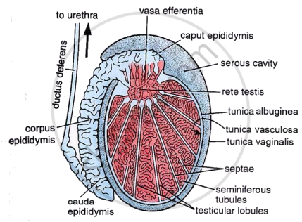
Internal morphology of testis
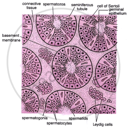
Testis in transverse section
CISCE: Class 10, 12
Key Points: Structure of Testis
- Each testis is enclosed by three protective layers – tunica vaginalis, tunica albuginea, and tunica vasculosa.
- Internally, each testis is divided into about 250 testicular lobules.
- Each lobule contains highly coiled seminiferous tubules, which are the site of spermatogenesis.
- The wall of seminiferous tubules is lined by germinal epithelium consisting of spermatogonia, Sertoli cells, and interstitial cells.
- Spermatogonia are diploid male germ cells that undergo meiosis to form haploid sperms.
- Sertoli cells act as nurse cells, providing nourishment and support to developing sperms, while Leydig cells secrete testosterone.
- Sperms move from seminiferous tubules into efferent ducts, then to the epididymis for maturation and temporary storage before entering the vas deferens.
