Topics
Reproduction in Lower and Higher Plants
- Reproduction
- Asexual Reproduction
- Asexual Reproduction in Unicellular Organisms > Binary Fission
- Binary Fission > Simple Binary Fission
- Binary Fission > Transverse Binary Fission
- Binary Fission > Longitudinal Binary Fission
- Asexual Reproduction in Unicellular Organisms > Multiple Fission
- Asexual Reproduction in Unicellular Organisms > Budding
- Asexual Reproduction in Multicellular Organisms > Fragmentation
- Asexual Reproduction in Multicellular Organisms > Spore Formation
- Vegetative Reproduction or Vegetative Propagation
- Natural Vegetative Propagation
- Artificial Vegetative Propagation
- Sexual Reproduction
- Flower
- Structure and Development of Anther
- Microsporogenesis
- Structure and Development of Male Gametophyte
- Structure and Development of Ovule
- Types of Ovules (Based on Orientation)
- Types of Ovules (Based on Integuments)
- Megasporogenesis
- Development of Female Gametophyte or Embryo Sac
- Pollination
- Autogamy
- Cross-pollination
- Geitonogamy
- Agents and Types of Cross-pollination
- Anemophily
- Hydrophily
- Entomophily
- Ornithophily
- Cheiropteriphily
- Malacophily
- Outbreeding Devices
- Fertilization
- Pollen Pistil Interaction
- Artificial Hybridization or Artificial Fertilization
- Double Fertilization and Triple Fusion
- Endosperm
- Embryo
- Seed and Fruit Development
- Dormancy
- Apomixis
- Parthenocarpy
- Polyembryony
- Overview of Reproduction in Lower and Higher Plants
Reproduction in Lower and Higher Animals
- Reproduction
- Asexual Reproduction
- Asexual Reproduction in Animals > Gemmule Formation
- Asexual Reproduction in Multicellular Organisms > Budding
- Asexual Reproduction in Multicellular Organisms > Regeneration
- Sexual Reproduction
- Human Reproduction
- The Male Reproductive System
- Basic Concept of Testes
- Duct system of Male Reproductive Tract
- Accessory Glands of Male Reproductive System
- Semen (Seminal fluid)
- External Genitalia: Penis
- The Female Reproductive System
- Ovaries
- Female Reproductive Duct System
- External Genitalia: Vulva
- Mammary Glands
- Puberty
- Menstrual Cycle (Ovarian Cycle)
- Major Events of Menstrual Cycle
- Menstrual Hygiene
- Gametogenesis
- Spermatogenesis
- Structure of Sperm
- Oogenesis
- Structure of Secondary Oocyte
- Fertilisation in Human
- Embryonic Development in Human
- Fate of Germ Layers in Embryonic Development
- Stem Cells
- Pregnancy in Humans
- Placenta (Growth) in Human
- Parturition (Birth) in Human
- Lactation in Human
- Concept of Reproductive Health
- Birth Control
- Natural Contraceptive Methods
- Artificial Contraceptive Methods
- Amniocentesis
- Sexually Transmitted Diseases (STD) or Sexually Transmitted Infections (STI)
- Infertility
- Assisted Reproductive Technology (ART)
- Overview of Reproduction in Lower and Higher Animals
Inheritance and Variation
- Heredity
- Gregor Johann Mendel – Father of Genetics
- Mendel's Experiments on Inheritance
- Reasons for Mendel's Success
- Genetic Terminology
- Monohybrid Cross
- Dihybrid Cross
- Mendel's Laws > The Law of Dominance
- Mendel's Laws > The Law of Segregation (Law of Purity of Gametes)
- Mendel's Laws > The Law of Independent Assortment
- Back Cross and Test Cross
- Deviations from Mendel’s Findings
- Exceptions to Mendel's Principles > Incomplete Dominance
- Exceptions to Mendel's Principles > Co-Dominance
- Exceptions to Mendel's Principles > Multiple alleles
- Exceptions to Mendel's Principles > Pleiotropy
- Chromosomal Theory of Inheritance
- Chromosomes - The Carriers of Heredity
- Types of Chromosomes
- Linkage and Crossing Over
- Autosomal Inheritance
- Sex Linked Inheritance
- Colour blindness
- Haemophilia
- Sex Determination
- Sex Determination in Humans
- Sex Determination in Birds
- Sex Determination in Honey Bees
- Human Genetic Disorders
- Thalassemia
- Down's syndrome (Mongolian Idiocy)
- Turner's Syndrome
- Klinefelter's Syndrome
- Overview of Inheritance and Variation
Molecular Basis of Inheritance
- Deoxyribonucleic Acid (DNA)
- Griffith’s Experiment
- Avery, McCarty and MacLeod’s Experiment
- The Hershey-Chase Experiment
- Packaging of DNA Helix
- DNA Replication
- Meselson and Stahl’s Experiment
- Mechanism of DNA Replication
- Semi-Conservative Replication
- Protein Synthesis
- Transcription
- Transcription Unit and the Gene
- Genetic Code
- Characteristics of the Genetic Code
- Mutations and Genetic Code
- tRNA – the Adapter Molecule
- Translation
- Mechanism of Translation
- Regulation of Gene Expression
- Operon Concept
- The Lac Operon
- Genomics
- Human Genome Project
- DNA Fingerprinting
- Overview of Molecular Basis of Inheritance
Origin and Evolution of Life
- Origin of Life on Earth
- Redi's and Louis Pasteur’s Experiment
- Chemical Evolution of Life
- Urey and Miller’s Experiment
- The RNA World
- Organic Evolution
- Darwin’s Theory of Natural Selection (Darwinism)
- Basic Postulates of Darwinism
- Drawbacks and Criticism of Darwinism
- Mutation Theory
- Modern Synthetic Theory of Evolution
- Modern Synthetic Theory of Evolution > Genetic Variations
- Modern Synthetic Theory of Evolution > Natural Selection
- Modern Synthetic Theory of Evolution > Isolation
- Mechanism of Organic Evolution
- Hardy Weinberg’s Principle
- Adaptive Radiation
- Evidences of Organic Evolution
- Evidences of organic evolution > Palaeontology
- Connecting Links
- Homology and Homologous Organs
- Analogy and Analogous Organs
- Vestigial Organs
- Molecular (Genetic) Evidences
- Speciation
- Geological Time Scale
- Human Evolution
- Stages of Human Evolution
- Overview of Origin and Evolution of Life
Plant Water Relation
- Properties of Water
- Water Absorbing Organ
- Water Available to Roots for Absorption
- Diffusion
- Osmosis
- Imbibition
- Osmotic Pressure
- Water Potential (ψ)
- Turgidity and Flaccidity (Plasmolysis)
- Path of Water Across the Root
- Mechanism of Absorption of Water
- Translocation of Water
- Root Pressure Theory (Vital Theory)
- Capillarity Theory (physical force theory)
- Cohesion-Tension Theory (Transpiration pull theory)
- Transport of Mineral Ions
- Transportation of Food and Other Substances
- Concept of Transpiration
- Types of Transpiration > Cuticular Transpiration
- Types of Transpiration > Lenticular Transpiration
- Types of Transpiration > Stomatal Transpiration
- Structure of Stomatal Apparatus
- Significance of Transpiration
- Overview of Plant Water Relation
Plant Growth and Mineral Nutrition
- Plant Growth
- Phases of Plant Growth
- Conditions for Plant Growth
- Plant Growth Rate
- Types of Plant Growth
- Plant Growth Curve
- Differentiation, De-differentiation, Re- Differentiation
- Plant Development
- Plant Plasticity
- Plant Hormones
- Auxins
- Gibberellins
- Cytokinins
- Ethylene
- Abscisic Acid (ABA)
- Photoperiodism
- Vernalization (Yarovization)
- Plant Mineral Nutrition
- Roles of Mineral Elements in Plants
- Minerals Salt Absorption
- Nitrogen Cycle
- Overview of Plant Growth and Mineral Nutrition
Respiration and Circulation
- Respiration
- Gaseous Exchange in plants
- Respiration in Animals
- Human Respiratory System
- Mechanism of Respiration > Breathing
- Mechanism of Respiration > External Respiration
- Mechanism of Respiration > Internal Respiration
- Cellular Respiration
- Regulation of Breathing / Respiration
- Disorders of Respiratory System
- Transportation in Living Organisms
- Circulation in Animals
- Circulatory System Or Blood Vascular System
- Composition of Blood > Cellular Elements: Red Blood Cells (Erythrocytes)
- Composition of Blood > Cellular Elements: White Blood Cells (Leukocytes)
- Composition of Blood > Cellular Elements: Blood Platelets (Thrombocytes)
- Human Heart
- Working Mechanism of Human Heart
- Blood Vessels
- Blood Pressure (B.P.)
- Electrocardiogram (ECG)
- Lymph and Lymphatic System
- Overview of Respiration and Circulation
Control and Co-ordination
- Need for Control and Coordination in Organisms
- Nervous System in Hydra
- Nervous System in Planaria (Flatworm)
- Neural Tissue
- Synapse
- Transmission and Generation of Nerve Impulse
- Central Nervous System (CNS)
- The Human Brain
- The Spinal Cord
- Peripheral Nervous System (PNS)
- Reflex Action
- Autonomic Nervous System (ANS)
- Sensory Receptors
- Human Eye
- Structure of the Eyeball
- Human Ear
- Internal Ear and the Mechanism of Balance
- Disorders of Nervous System
- Human Endocrine System
- Concept of Hormone
- General Properties of Hormones
- Mechanism of Hormone Action
- The Hypothalamus
- Pituitary Gland or Hypophysis Gland
- The Pineal Gland
- Thyroid Gland
- Parathyroid Gland
- Thymus Gland
- Adrenal Gland (Suprarenal Gland)
- Pancreas (Islets of Langerhans)
- Reproductive Glands (Gonads)
- Diffuse Endocrine Glands
- Overview of Control and Co-ordination
Human Health and Diseases
- Health
- The Immune System
- Immunity
- Types of Immunity > Innate Immunity
- Types of Immunity > Acquired Immunity
- Cells of Immune System
- Vaccination and Immunization
- Structure of Antibody
- Formation of Antigen-Antibody Complex
- Blood Transfusion and Blood Groups (ABO and Rh system)
- Disease
- Diseases Caused by Protozoa > Malaria
- Diseases Caused by Protozoa > Amoebiasis (Amoeboic dysentery)
- Diseases Caused by Helminths > Ascariasis
- Diseases Caused by Helminths > Filariasis (Elephantiasis)
- Diseases Caused by Bacteria > Typhoid
- Diseases Caused by Bacteria > Pneumonia
- Diseases Caused by Viruses > Common Cold
- Diseases Caused by Fungi > Ringworm
- Diseases Caused by Viruses > Dengue Fever
- Types of Cancer
- Causes of Cancer
- Organs Commonly Affected by Cancer
- Prevention/Treatment of Cancer
- Acquired Immuno Deficiency Syndrome (AIDS)
- Concept of Adolescence
- Addiction
- Drug Abuse
- Addiction and Dependence
- Effects of Drug and Alcohol Abuse
- Prevention and Control of Drugs and Alcohol Abuse
- Cancer
Human Reproduction
Enhancement of Food Production
- Improvement in Food Production
- Plant Breeding
- Hybridization and its Technique
- Mutation Breeding
- Tissue Culture
- Single Cell Protein (SCP)
- Biofortification
- Animal Husbandry (Livestock)
- Animal Husbandry (Livestock) > Animal Breeding
- Animal Husbandry (Livestock) > Dairy (Livestock) Farm Management
- Animal Husbandry (Livestock) > Poultry Farm Management
- Animal Husbandry (Livestock) > Apiculture (Bee Farming)
- Animal Husbandry (Livestock) > Pisciculture (Fish Farming)
- Animal Husbandry (Livestock) > Sericulture
- Animal Husbandry (Livestock) > Lac Culture
- Microbes in Human Welfare
- Microbes in Industrial Products
- Microbes in Sewage Treatment
- Microbes in Energy Generation
- Microbes as Biocontrol Agents
- Microbes as Biofertilizers
- Microbial Role in Dairy Products
- Overview of Enhancement of Food Production
Biotechnology
- Concept of Biotechnology
- Principles of Processes of Biotechnology
- Technique of Gene Cloning and rDNA Technology
- Methodology for rDNA Technology
- Applications of Biotechnology in Health and Medicine
- Applications of Biotechnology in Agriculture
- Gene Therapy
- Crop Biotechnology > Genetically Modified Organisms (GMOs)
- Transgenic Plants
- Transgenic Animals
- Bioethics
- Effects of Biotechnology on the Environment
- Effects of Biotechnology on Human Health
- Biopatent
- Biopiracy
- Overview of Biotechnology
Organisms and Populations
- Organisms and Their Environment
- Habitat
- Niche
- Habitat Vs Niche
- Climatic Factors > Temperature
- Climatic Factors > Precipitation
- Climatic Factors > Light
- Soil Factors (Edaphic Factors)
- Adaptations
- Population
- Population Age Distribution
- Population Growth
- Population Interactions
- Positive Interactions > Mutualism (Symbiosis)
- Negative Interactions > Competition
- Negative Interactions > Parasitism
- Negative Interactions > Predation
- Positive Interactions > Commensalism
- Overview of Organisms and Populations
Ecosystems and Energy Flow
Biodiversity, Conservation and Environmental Issues
- Biodiversity
- Levels of Biodiversity
- Patterns of Biodiversity
- Importance of Species Diversity to the Ecosystem
- Biodiversity Current Scenario
- Loss of Biodiversity
- Threatened Species
- Conservation of Biodiversity
- Biodiversity Conservation Methods
- Biological Diversity Act, 2002
- Environmental Issues
- Air Pollution
- Effects of Air Pollution
- Major Air Pollutants
- Prevention and Control of Air Pollution
- Noise Pollution
- Water Pollution
- Sources of Water Pollution
- Prevention and Control of Water Pollution
- Solid Waste Management
- Greenhouse Effect
- Global Warming
- Ozone Layer Depletion
- Deforestation and Its Causes
- Mission Harit Maharashtra
- Overview of Biodiversity, Conservation and Environmental Issues
Maharashtra State Board: Class 12
CISCE: Class 12
Definition: Embryo
The embryo is the young developing plant formed from the zygote after fertilisation. It develops at the micropylar end of the embryo sac.
Maharashtra State Board: Class 12
CISCE: Class 12
Structure of the Dicot Embryo
A typical dicotyledonous embryo consists of an embryonal axis and two cotyledons attached laterally to it. The embryonal axis has distinct upper and lower regions:
| Part | Location on Embryonal Axis | Terminates In / Function |
|---|---|---|
| Epicotyl | Above the cotyledons | Terminates in the plumule (shoot tip / stem tip) |
| Hypocotyl | Below the cotyledons (cylindrical portion) | Terminates in the radicle (root tip) |
| Radicle | Lowermost end | Develops into the primary root |
| Root Cap (Calyptra) | Covers the radicle tip | Protects the root tip during soil penetration |
| Plumule | Apical shoot tip | Develops into the shoot system; first leaves emerge from here |
| Cotyledons (×2) | Attached laterally to embryonal axis | Store food; function as first seed-leaves |
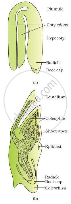
(a) A typical dicot embryo; (b) L.S. of an embryo of grass
Maharashtra State Board: Class 12
CISCE: Class 12
Step-by-Step Development - Dicot Embryo (Capsella)
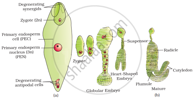
(a) Fertilised embryo sac showing zygote and Primary Endosperm Nucleus (PEN); (b) Stages in embryo development in a dicot [shown in reduced size as compared to (a)]
The following stages describe embryogenesis in Capsella bursa-pastoris:
The zygote divides transversely into two unequal cells:
- Basal cell (larger, towards micropyle) → will form the suspensor
- Terminal / Apical cell (smaller, towards chalaza) → will form the embryo proper
The basal cell divides transversely to form a filamentous suspensor of 6–10 cells.
- The first cell of the suspensor (micropylar end) becomes swollen and functions as a haustorium — it absorbs nutrients from the surrounding endosperm and passes them to the embryo.
- The lowermost cell of the suspensor is the hypophysis — this later gives rise to part of the radicle and root cap.
The terminal (embryonal initial) cell undergoes three successive mitotic divisions. Planes of division are at right angles to each other, yielding 8 cells arranged in two tiers:
- Upper tier (4 cells) → forms the plumule and cotyledons
- Lower tier (4 cells) → forms the hypocotyl and radicle
Periclinal divisions (parallel to the surface) of the outer cells of the octant produce a distinct outermost layer called the dermatogen, which will become the future epidermis. The embryo now takes on a spherical (globular) shape. The inner regions differentiate into procambium and ground meristem.
Cells of the upper tier begin to divide unevenly. Two lateral lobes (future cotyledons) grow rapidly, giving the embryo a characteristic heart shape. The terminal plumule and cotyledon initials become clearly visible.
Enlargement of the hypocotyl and further growth of the two cotyledons causes the embryo to curve. The final shape is horseshoe-shaped (U-shaped), characteristic of a mature dicot embryo. The suspensor degenerates at this stage.
Maharashtra State Board: Class 12
CISCE: Class 12
Structure of the Monocot Embryo
In monocotyledonous plants (e.g., the grass family), the embryo possesses only one cotyledon situated towards one side (laterally) of the embryonal axis. This single cotyledon is called the scutellum.
Two specialised protective sheaths are present — one for the shoot and one for the root — which are unique to monocots:
| Part | Location | Function |
|---|---|---|
| Scutellum | Lateral side of embryonal axis (one cotyledon) | Absorbs nutrients from the endosperm during germination; shield-shaped |
| Epicotyl | Above scutellum attachment point | Bears the shoot apex |
| Coleoptile | Hollow, foliar sheath enclosing the epicotyl | Protects the shoot (plumule + leaf primordia) during germination and soil emergence |
| Epiblast | Opposite side of the scutellum | Rudimentary / vestigial structure (considered vestigial second cotyledon) |
| Radicle | Lower end of embryonal axis | Gives rise to the primary root |
| Root Cap | Covering the radicle tip | Protects the root tip |
| Coleorhiza | Undifferentiated sheath surrounding the radicle + root cap | Protects the root (radicle) during germination |
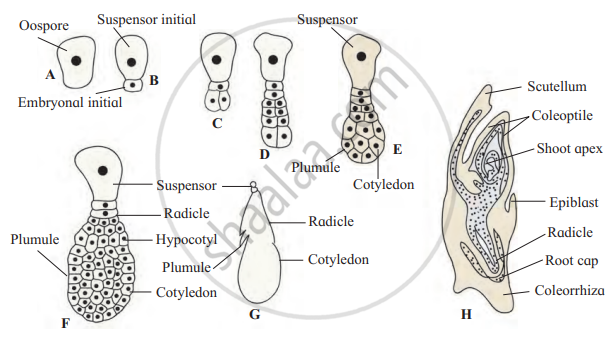
Development of Monocot (grass) Embryo
Maharashtra State Board: Class 12
CISCE: Class 12
Key Points: Embryo
- Embryogenesis is the process of development of the zygote into an embryo, which begins after fertilisation and some endosperm formation.
- The embryo develops at the micropylar end of the embryo sac.
- The zygote divides to form a two-celled proembryo: a terminal (embryonal) cell and a basal (suspensor) cell.
- The suspensor pushes the developing embryo into the endosperm and helps in nutrient supply, while the embryonal cell forms the main embryo.
- The embryo passes through stages like the octant and heart-shaped stage, forming structures such as cotyledons, plumule, radicle, and hypocotyl.
- In monocots, a single cotyledon (scutellum) is present with protective sheaths, the coleoptile (plumule) and coleorhiza (radicle), while in dicots, two cotyledons are formed.
