Advertisements
Advertisements
प्रश्न
Which of the given statements is correct in the context of visualizing DNA molecules separated by agarose gel electrophoresis?
पर्याय
DNA can be seen in visible light
DNA can be seen without staining in visible light
Ethidium bromide stained DNA can be seen in visible light
Ethidium bromide stained DNA can be seen under exposure to UV light
Advertisements
उत्तर
Ethidium bromide stained DNA can be seen under exposure to UV light
Explanation:
DNA molecules separated by agarose gel electrophoresis are not visible in normal visible light and require staining. Ethidium bromide binds to DNA and fluoresces under UV light, allowing visualization of DNA bands as bright orange coloured bands. This method is widely used in laboratories for DNA analysis after gel electrophoresis.
APPEARS IN
संबंधित प्रश्न
Explain with the help of a suitable example the naming of a restriction endonuclease.
How are 'sticky ends' formed on a DNA strand? Why are they so called?
Why is the enzyme cellulase needed for isolating genetic material from plant cells and not form the animal cells?
Mention the difference in the mode of action of exonuclease and endonuclease.
Suggest a technique to a researcher who needs to separate fragments of DNA.
How does a restriction nuclease function? Explain
Make a chart (with diagrammatic representation) showing a restriction enzyme, the substrate DNA on which it acts, the site at which it cuts DNA and the product it produces.
Collect 5 examples of palindromic DNA sequences. Better try to create a palindromic sequence by following base-pair rules.
Explain briefly:
Restriction enzymes and DNA
Distinguish between exonuclease and endonuclease.
How does restriction endonuclease function?
Which of the following radioisotope is not suitable for DNA labeling based studies?
'Restriction' in Restriction enzyme refers to ______.
While isolating DNA from bacteria, which of the following enzymes is not required?
Which of the following bacteria is not a source of restriction endonuclease?
CTTAAG
GAATTC
- What are such sequences called? Name the enzyme used that recognizes such nucleotide sequences.
- What is their significance in biotechnology?
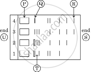
Given below is the stepwise schematic representation of the process of electrophoresis. Identify the 'alphabets' representing
- Anode end
- smallest/lightest DNA strand in the matrix
- Agarose gel
What are the protruding and hanging stretches of DNA produced by these restriction enzymes called? Describe their role in the formation of rDNA.
Identify the activity of endonuclease and exonuclease in the given image.
Which of the following was the very first restriction endonuclease to be isolated and characterized?
How do exonucleases differ fundamentally from endonucleases in their enzymatic site of action?
In the standard nomenclature convention for restriction endonucleases, what do the second and third letters signify?
Which of the following represents the specific palindromic recognition sequence cleaved by the restriction endonuclease EcoRI?
During agarose gel electrophoresis, why do DNA fragments migrate toward the anode (positive electrode)?
What is the origin of agarose, the matrix most commonly used in gel electrophoresis?
How are separated DNA bands visualized following agarose gel electrophoresis?
Why are Type II restriction endonucleases the primary class of restriction enzymes used in genetic manipulation?
