Advertisements
Advertisements
प्रश्न
How are DNA fragments visualised once they are separated by gel electrophoresis?
Advertisements
उत्तर
The separated DNA fragments can be visualised only after staining the DNA with a compound known as ethidium bromide followed by exposure to UV radiation (you cannot see pure DNA fragments in the visible light and without staining). You can see bright orange coloured bands of DNA in a ethidium bromide stained gel exposed to UV light.
APPEARS IN
संबंधित प्रश्न
Explain briefly:
Restriction enzymes and DNA
Answer the following question.
Explain the significance of palindromic nucleotide sequence in the formation of recombinant DNA.
DNA fragments separate according to size through?
In agarose gel electrophoresis, DNA molecules are separated on the basis of their ______.
While isolating DNA from bacteria, which of the following enzymes is not required?
The role of DNA ligase in the construction of a recombinant DNA molecule is ______.
Restriction enzymes that are used in the construction of recombinant DNA are endonucleases which cut the DNA at ‘specific-recognition sequence’. What would be the disadvantage if they do not cut the DNA at specific-recognition sequence?
How does one visualise DNA on an agarose gel?
CTTAAG
GAATTC
- What are such sequences called? Name the enzyme used that recognizes such nucleotide sequences.
- What is their significance in biotechnology?
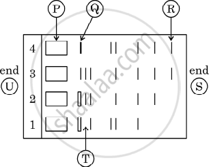
Given below is the stepwise schematic representation of the process of electrophoresis. Identify the 'alphabets' representing
- Anode end
- smallest/lightest DNA strand in the matrix
- Agarose gel