Advertisements
Advertisements
प्रश्न
Explain briefly:
Restriction enzymes and DNA
Advertisements
उत्तर
Restriction enzymes belong to a larger class of enzymes called nucleases. They are mainly classified into two types:
- Exonucleases: They remove nucleotides from the terminal ends of the DNA molecule.
- Endonucleases: They make cuts at specific positions anywhere within the DNA molecule.
Restriction endonucleases are a specific class of endonucleases that recognise palindromic nucleotide sequences and cleave DNA at specific sites. They cut both strands of the DNA double helix at specific points to generate sticky or blunt ends, which are essential for creating recombinant DNA.
APPEARS IN
संबंधित प्रश्न
How are 'sticky ends' formed on a DNA strand? Why are they so called?
Why is the enzyme cellulase needed for isolating genetic material from plant cells and not form the animal cells?
Suggest a technique to a researcher who needs to separate fragments of DNA.
How does a restriction nuclease function? Explain
Name and describe the technique that helps in separating the DNA fragments formed by the use of restriction endonuclease
Make a chart (with diagrammatic representation) showing a restriction enzyme, the substrate DNA on which it acts, the site at which it cuts DNA and the product it produces.
Distinguish between exonuclease and endonuclease.
Explain the roles of the following with the help of an example each in recombinant DNA technology :
Restriction Enzymes
Answer the following question.
Explain the significance of palindromic nucleotide sequence in the formation of recombinant DNA.
Answer the following question.
Write the use of restriction endonuclease in the formation of recombinant DNA.
The total number of nucleotide sequences of DNA that code for a hormone is 1530. The proportion of different bases in the sequence is found to be Adenine = 34%, Guanine = 19%, Cytosine = 23%, Thymine = 19%.
Applying Chargaff’s rule, what conclusion can be drawn?
Which of the following radioisotope is not suitable for DNA labeling based studies?
Restriction enzymes ______.
A specific recognition sequence identified by endonucleases to make cuts at specific positions within the DNA is ______
In agarose gel electrophoresis, DNA molecules are separated on the basis of their ______.
Which of the following statements does not hold true for restriction enzyme?
Would you choose an exonuclease while producing a recombinant DNA molecule?
What does H in’ ‘d’ and ‘III’ refer to in the enzyme Hind III?
Restriction enzymes should not have more than one site of action in the cloning site of a vector. Comment.
Restriction enzymes that are used in the construction of recombinant DNA are endonucleases which cut the DNA at ‘specific-recognition sequence’. What would be the disadvantage if they do not cut the DNA at specific-recognition sequence?
A mixture of fragmented DNA was electrophoresed in an agarose gel. After staining the gel with ethidium bromide, no DNA bands were observed. What could be the reason?
CTTAAG
GAATTC
- What are such sequences called? Name the enzyme used that recognizes such nucleotide sequences.
- What is their significance in biotechnology?
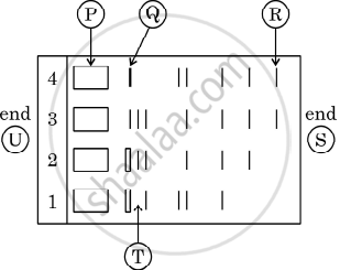
Given below is the stepwise schematic representation of the process of electrophoresis. Identify the 'alphabets' representing
- Anode end
- smallest/lightest DNA strand in the matrix
- Agarose gel
What is elution?
Given below is the restriction site of a restriction endonuclease Pst-I and the cleavage sites on a DNA molecule.
\[\ce{5' C - T - G - C - A \overset{\downarrow}{-}{G 3'}}\]
\[\ce{3' G\underset{\uparrow}{-} A - C - G - T - C 5'}\]
Choose the option that gives the correct resultant fragments by the action of the enzyme Pst-I.
'EcoRI' has played a very significant role in rDNA technology.
- Explain the convention for naming EcoRI.
- Write the recognition site and the cleavage sites of this restriction endonuclease.
State the principle involved in separation of DNA fragments using gel electrophoresis.
Identify the activity of endonuclease and exonuclease in the given image.
