Advertisements
Advertisements
प्रश्न
Carefully observe the given picture. A mixture of DNA with fragments ranging from 200 base pairs to 2500 base pairs was electrophoresed on agarose gel with the following arrangement.
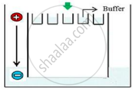
(a) What result will be obtained on staining with ethidium bromide? Explain with reason.
(b) The above setup was modified and a band with 250 base pairs was obtained at X.
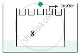
What change(s) were made to the previous design to obtain a band at X? Why did the band appear at position X?
Advertisements
उत्तर
(a) No bands will be obtained as all DNA will be seen in the well only; DNA fragments being negatively charged will not move towards the negative end/cathode. DNA being negatively charged will remain stationed at the positive end/anode end of the agar block.
(b)
- The position of the positive terminal/end/anode and the negative terminal/end/cathode was inter-changed.
- The fragment with the least base pairs will get separated faster and move faster to the anode end.
APPEARS IN
संबंधित प्रश्न
How does a restriction nuclease function? Explain
Collect 5 examples of palindromic DNA sequences. Better try to create a palindromic sequence by following base-pair rules.
Give a reason why :
Single cloning site is preferred in a vector.
The total number of nucleotide sequences of DNA that code for a hormone is 1530. The proportion of different bases in the sequence is found to be Adenine = 34%, Guanine = 19%, Cytosine = 23%, Thymine = 19%.
Applying Chargaff’s rule, what conclusion can be drawn?
Molecular scissors, which cut DNA at specific site is ______.
Which of the following enzymes catalyse the removal of nucleotides from the ends of DNA?
Which of the given statements is correct in the context of visualizing DNA molecules separated by agarose gel electrophoresis?
Which of the following statements does not hold true for restriction enzyme?
Would you choose an exonuclease while producing a recombinant DNA molecule?
Hind II always cuts DNA molecules at a particular point called recognition sequence and it consists of ______.
