Advertisements
Advertisements
प्रश्न
A mixture of fragmented DNA was electrophoresed in an agarose gel. After staining the gel with ethidium bromide, no DNA bands were observed. What could be the reason?
Advertisements
उत्तर
The reasons are as follows:
- DNA sample that was loaded on the gel may have got contaminated with nuclease (exo-or endo-or both) and completely degraded.
- Electrodes were put in opposite orientation in the gel assembly that is anode towards the wells (where DNA sample is loaded). Since DNA molecules are negatively charged, they move towards anode and hence move out of the gel instead of moving into the matrix of gel.
- Ethidium bromide was not added at all or was not added in sufficient concentration and DNA was not visible.
APPEARS IN
संबंधित प्रश्न
Explain with the help of a suitable example the naming of a restriction endonuclease.
Why is the enzyme cellulase needed for isolating genetic material from plant cells and not form the animal cells?
Make a chart (with diagrammatic representation) showing a restriction enzyme, the substrate DNA on which it acts, the site at which it cuts DNA and the product it produces.
Explain the roles of the following with the help of an example each in recombinant DNA technology :
Restriction Enzymes
There is a restriction endonudease called as EcoRI. What does co part in it stands for?
DNA fragments separate according to size through?
DNA strands on a gel stained with ethidium bromide when viewed under UV radiation, appear as ______
In agarose gel electrophoresis, DNA molecules are separated on the basis of their ______.
Restriction enzymes that are used in the construction of recombinant DNA are endonucleases which cut the DNA at ‘specific-recognition sequence’. What would be the disadvantage if they do not cut the DNA at specific-recognition sequence?
How does one visualise DNA on an agarose gel?
Carefully observe the given picture. A mixture of DNA with fragments ranging from 200 base pairs to 2500 base pairs was electrophoresed on agarose gel with the following arrangement.
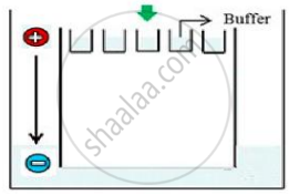
(a) What result will be obtained on staining with ethidium bromide? Explain with reason.
(b) The above setup was modified and a band with 250 base pairs was obtained at X.
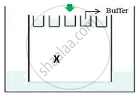
What change(s) were made to the previous design to obtain a band at X? Why did the band appear at position X?
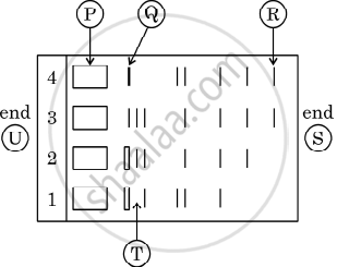
Given below is the stepwise schematic representation of the process of electrophoresis. Identify the 'alphabets' representing
- Anode end
- smallest/lightest DNA strand in the matrix
- Agarose gel
What are the protruding and hanging stretches of DNA produced by these restriction enzymes called? Describe their role in the formation of rDNA.
State the principle involved in separation of DNA fragments using gel electrophoresis.
How are DNA fragments visualised once they are separated by gel electrophoresis?
Identify the activity of endonuclease and exonuclease in the given image.
Hind II always cuts DNA molecules at a particular point called recognition sequence and it consists of ______.
