Advertisements
Advertisements
Question
How does one visualise DNA on an agarose gel?
Advertisements
Solution
A compound called Ethidium Bromide stains DNA, which on irradiating with Ultraviolet, fluoresce gives orange light. Hence, DNA fragments appear as orange band in the presence of Ethidium Bromide and UV.
APPEARS IN
RELATED QUESTIONS
How are 'sticky ends' formed on a DNA strand? Why are they so called?
How does a restriction nuclease function? Explain
Make a chart (with diagrammatic representation) showing a restriction enzyme, the substrate DNA on which it acts, the site at which it cuts DNA and the product it produces.
Explain briefly:
Restriction enzymes and DNA
Distinguish between exonuclease and endonuclease.
How does restriction endonuclease function?
The total number of nucleotide sequences of DNA that code for a hormone is 1530. The proportion of different bases in the sequence is found to be Adenine = 34%, Guanine = 19%, Cytosine = 23%, Thymine = 19%.
Applying Chargaff’s rule, what conclusion can be drawn?
Molecular scissors, which cut DNA at specific site is ______.
Which of the following enzymes catalyse the removal of nucleotides from the ends of DNA?
What does H in’ ‘d’ and ‘III’ refer to in the enzyme Hind III?
Restriction enzymes should not have more than one site of action in the cloning site of a vector. Comment.
A plasmid DNA and a linear DNA (both are of the same size) have one site for a restriction endonuclease. When cut and separated on agarose gel electrophoresis, plasmid shows one DNA band while linear DNA shows two fragments. Explain.
CTTAAG
GAATTC
- What are such sequences called? Name the enzyme used that recognizes such nucleotide sequences.
- What is their significance in biotechnology?
Carefully observe the given picture. A mixture of DNA with fragments ranging from 200 base pairs to 2500 base pairs was electrophoresed on agarose gel with the following arrangement.
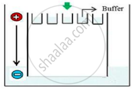
(a) What result will be obtained on staining with ethidium bromide? Explain with reason.
(b) The above setup was modified and a band with 250 base pairs was obtained at X.
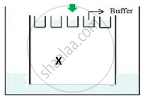
What change(s) were made to the previous design to obtain a band at X? Why did the band appear at position X?
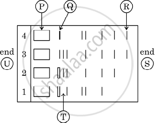
Given below is the stepwise schematic representation of the process of electrophoresis. Identify the 'alphabets' representing
- Anode end
- smallest/lightest DNA strand in the matrix
- Agarose gel
Given below is the restriction site of a restriction endonuclease Pst-I and the cleavage sites on a DNA molecule.
\[\ce{5' C - T - G - C - A \overset{\downarrow}{-}{G 3'}}\]
\[\ce{3' G\underset{\uparrow}{-} A - C - G - T - C 5'}\]
Choose the option that gives the correct resultant fragments by the action of the enzyme Pst-I.
What are the protruding and hanging stretches of DNA produced by these restriction enzymes called? Describe their role in the formation of rDNA.
During agarose gel electrophoresis, why do DNA fragments migrate toward the anode (positive electrode)?
