Advertisements
Advertisements
Question
A plasmid DNA and a linear DNA (both are of the same size) have one site for a restriction endonuclease. When cut and separated on agarose gel electrophoresis, plasmid shows one DNA band while linear DNA shows two fragments. Explain.
Advertisements
Solution
It is because plasmid is a circular DNA molecule. When cut with enzyme, it becomes linear but does not get fragmented. Whereas, a linear DNA molecule gets cut into two fragments. Hence, a single DNA band is observed for plasmid while two DNA bands are observed for linear DNA in agarose gel.

APPEARS IN
RELATED QUESTIONS
Mention the difference in the mode of action of exonuclease and endonuclease.
Suggest a technique to a researcher who needs to separate fragments of DNA.
Name and describe the technique that helps in separating the DNA fragments formed by the use of restriction endonuclease
Explain briefly:
Restriction enzymes and DNA
Distinguish between exonuclease and endonuclease.
How does restriction endonuclease function?
Answer the following question.
Write the use of restriction endonuclease in the formation of recombinant DNA.
Molecular scissors, which cut DNA at specific site is ______.
'Restriction' in restriction enzyme refers to
DNA strands on a gel stained with ethidium bromide when viewed under UV radiation, appear as ______
Which of the following enzymes catalyse the removal of nucleotides from the ends of DNA?
In agarose gel electrophoresis, DNA molecules are separated on the basis of their ______.
Which of the following statements does not hold true for restriction enzyme?
What does H in’ ‘d’ and ‘III’ refer to in the enzyme Hind III?
How does one visualise DNA on an agarose gel?
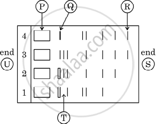
Given below is the stepwise schematic representation of the process of electrophoresis. Identify the 'alphabets' representing
- Anode end
- smallest/lightest DNA strand in the matrix
- Agarose gel
What is elution?
State the principle involved in separation of DNA fragments using gel electrophoresis.
