Advertisements
Advertisements
Question
Which of the following bacteria is not a source of restriction endonuclease?
Options
Haemophilus influenzae
Escherichia coli
Entamoeba coli
Bacillus amyloliquefaciens
Advertisements
Solution
Entamoeba coli
APPEARS IN
RELATED QUESTIONS
Why is the enzyme cellulase needed for isolating genetic material from plant cells and not form the animal cells?
Mention the difference in the mode of action of exonuclease and endonuclease.
How does a restriction nuclease function? Explain
Collect 5 examples of palindromic DNA sequences. Better try to create a palindromic sequence by following base-pair rules.
Explain the roles of the following with the help of an example each in recombinant DNA technology :
Restriction Enzymes
A mixture containing DNA fragments a, b, c and d, with molecular weights of a + b = c, a > b and d > c was subject to agarose get electrophoresis. This position of these fragments from cathode to anode to anode sides of the gel would be ______.
Which of the following radioisotope is not suitable for DNA labeling based studies?
There is a restriction endonudease called as EcoRI. What does co part in it stands for?
Molecular scissors, which cut DNA at specific site is ______.
DNA strands on a gel stained with ethidium bromide when viewed under UV radiation, appear as ______
Which of the given statements is correct in the context of visualizing DNA molecules separated by agarose gel electrophoresis?
While isolating DNA from bacteria, which of the following enzymes is not required?
The role of DNA ligase in the construction of a recombinant DNA molecule is ______.
Restriction enzymes should not have more than one site of action in the cloning site of a vector. Comment.
How does one visualise DNA on an agarose gel?
Carefully observe the given picture. A mixture of DNA with fragments ranging from 200 base pairs to 2500 base pairs was electrophoresed on agarose gel with the following arrangement.
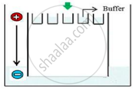
(a) What result will be obtained on staining with ethidium bromide? Explain with reason.
(b) The above setup was modified and a band with 250 base pairs was obtained at X.

What change(s) were made to the previous design to obtain a band at X? Why did the band appear at position X?
What is elution?
State the importance of elution in this process.
