Advertisements
Advertisements
प्रश्न
Which of the following bacteria is not a source of restriction endonuclease?
पर्याय
Haemophilus influenzae
Escherichia coli
Entamoeba coli
Bacillus amyloliquefaciens
Advertisements
उत्तर
Entamoeba coli
APPEARS IN
संबंधित प्रश्न
Suggest a technique to a researcher who needs to separate fragments of DNA.
Make a chart (with diagrammatic representation) showing a restriction enzyme, the substrate DNA on which it acts, the site at which it cuts DNA and the product it produces.
Collect 5 examples of palindromic DNA sequences. Better try to create a palindromic sequence by following base-pair rules.
Explain briefly:
Restriction enzymes and DNA
The DNA fragment separated on an agarose gel can be visualized by staining with ______.
Restriction enzymes ______.
There is a restriction endonudease called as EcoRI. What does co part in it stands for?
DNA strands on a gel stained with ethidium bromide when viewed under UV radiation, appear as ______
A specific recognition sequence identified by endonucleases to make cuts at specific positions within the DNA is ______
In agarose gel electrophoresis, DNA molecules are separated on the basis of their ______.
While isolating DNA from bacteria, which of the following enzymes is not required?
What does H in’ ‘d’ and ‘III’ refer to in the enzyme Hind III?
How does one visualise DNA on an agarose gel?
A mixture of fragmented DNA was electrophoresed in an agarose gel. After staining the gel with ethidium bromide, no DNA bands were observed. What could be the reason?
CTTAAG
GAATTC
- What are such sequences called? Name the enzyme used that recognizes such nucleotide sequences.
- What is their significance in biotechnology?
Carefully observe the given picture. A mixture of DNA with fragments ranging from 200 base pairs to 2500 base pairs was electrophoresed on agarose gel with the following arrangement.
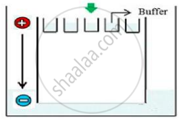
(a) What result will be obtained on staining with ethidium bromide? Explain with reason.
(b) The above setup was modified and a band with 250 base pairs was obtained at X.
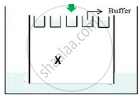
What change(s) were made to the previous design to obtain a band at X? Why did the band appear at position X?
State the importance of elution in this process.
State the principle involved in separation of DNA fragments using gel electrophoresis.
