Topics
Reproduction in Organisms
Sexual Reproduction in Flowering Plants
- Flower
- Pre-fertilisation in Flowering Plant: Structures and Events
- Structure and Development of Anther
- Microsporogenesis
- Structure and Development of Male Gametophyte
- Pollen Viability and Storage
- Structure and Development of Ovule
- Megasporogenesis
- Development of Female Gametophyte or Embryo Sac
- Pollination
- Autogamy
- Geitonogamy
- Cross-pollination
- Agents of Pollination
- Anemophily
- Hydrophily
- Animal-Mediated Pollination (Zoophily)
- Outbreeding Devices
- Pollen Pistil Interaction
- Artificial Hybridization or Artificial Fertilization
- Double Fertilization and Triple Fusion
- Events in Sexual Reproduction > Post-Fertilization Structures and Events
- Endosperm
- Dicotyledonous Embryo
- Monocotyledonous Embryo
- The Seed
- Apomixis
- Polyembryony
Reproduction
Genetics and Evolution
Human Reproduction
- Human Reproduction
- The Male Reproductive System
- The Female Reproductive System
- Gametogenesis
- Spermatogenesis
- Structure of Sperm
- Spermiogenesis
- Oogenesis
- Menstrual Cycle (Ovarian Cycle)
- Major Events of Menstrual Cycle
- Menstrual Hygiene
- Fertilization in Humans
- Implantation in Human
- Pregnancy and Embryonic Development
- Parturition (Birth) in Human
- Lactation in Human
Reproductive Health
- Concept of Reproductive Health
- Population Explosion and Control Measures
- Birth Control
- Natural Contraceptive Methods
- Artificial Contraceptive Methods
- Induced Abortion or Medical Termination of Pregnancy (MTP)
- Sexually Transmitted Diseases (STD) or Sexually Transmitted Infections (STI)
- Infertility
- Assisted Reproductive Technology (ART)
- Amniocentesis
- Genetic Counselling
Biology and Human Welfare
Biotechnology
Environmental Issues
- Controlling Vehicular Air Pollution: a Case Study of Delhi
- Effects of Domestic Sewage and Industrial Effluents on Water
- Solid Wastes
- Radioactive Wastes
- Greenhouse Effect and Climate Change
- Ozone Depletion in the Stratosphere
- Degradation by Improper Resource Utilisation and Maintenance
- Radioactive Waste Management and E-waste
Principles of Inheritance and Variation
- Heredity and Variation
- Gregor Johann Mendel – Father of Genetics
- Mendel's Experiments on Inheritance
- Monohybrid Cross
- Punnett Square
- Back Cross and Test Cross
- Mendel's Laws > The Law of Dominance
- Mendel's Laws > The Law of Segregation (Law of Purity of Gametes)
- Exceptions to Mendel's Principles > Incomplete Dominance
- Exceptions to Mendel's Principles > Co-Dominance
- Dihybrid Cross
- Mendel's Laws > The Law of Independent Assortment
- Chromosomal Theory of Inheritance
- Linkage and Recombination
- Polygenic Inheritance
- Exceptions to Mendel's Principles > Pleiotropy
- Sex Determination
- Sex Determination in Humans
- Sex Determination in Honey Bees
- Mutations
- Pedigree Analysis
- Mendelian Disorders in Humans
- Chromosomal Disorders or Abnormalities
Molecular Basis of Inheritance
- Deoxyribonucleic Acid (DNA)
- Structure of Polynucleotide Chain
- Packaging of DNA Helix
- Search for Genetic Material
- Griffith’s Experiment
- Avery, McCarty and MacLeod’s Experiment
- The Hershey-Chase Experiment
- Properties of Genetic Material
- The RNA World
- DNA Replication
- Conservative Replication
- Dispersive Replication
- Semi-Conservative Replication
- Meselson and Stahl’s Experiment
- Enzymes used in DNA Replication
- Mechanism of DNA Replication
- Protein Synthesis
- Reverse Transcription (Teminism)
- Transcription
- Transcription Unit and the Gene
- Process of Transcription in Bacteria
- Process of Transcription in Eukaryotes
- Genetic Code
- Characteristics of the Genetic Code
- Mutations and Genetic Code
- Transfer RNA (tRNA)
- Translation
- Regulation of Gene Expression
- The Lac Operon
- Human Genome Project
- DNA Fingerprinting
Ecology
Evolution
- Origin of Life on Earth
- Urey and Miller’s Experiment
- Evolution of Life Forms - a Theory
- Evidences Supporting the Theory of Evolution
- Adaptive Radiation
- Theories and Mechanism of Evolution
- Theories and Mechanism of Evolution
- Hardy Weinberg’s Principle
- Types of Selection
- Brief Account of Evolution
- Human Evolution
Human Health and Diseases
- Concept and Determinants of Health
- Modes of Transmission of Diseases through Pathogens
- Diseases Caused by Bacteria > Typhoid
- Diseases Caused by Bacteria > Pneumonia
- Diseases Caused by Viruses > Common Cold
- Diseases Caused by Protozoa > Malaria
- Diseases Caused by Protozoa > Amoebiasis (Amoeboic dysentery)
- Diseases Caused by Helminths > Ascariasis
- Diseases Caused by Helminths > Filariasis (Elephantiasis)
- Diseases Caused by Fungi > Ringworm
- Prevention and Control of Infectious Diseases
- Immunity
- Types of Immunity > Innate Immunity
- Types of Immunity > Acquired Immunity
- Vaccination and Immunization
- Allergies
- Autoimmunity
- The Immune System
- Acquired Immuno Deficiency Syndrome (AIDS)
- Cancer
- Causes of Cancer
- Symptoms and Diagnosis of Cancer
- Prevention/Treatment of Cancer
- Drugs and Alcohol Abuse
- Addiction and Dependence
- Effects of Drug and Alcohol Abuse
- Prevention and Control of Drugs and Alcohol Abuse
Microbes in Human Welfare
Strategies for Enhancement in Food Production
Biotechnology - Principles and Processes
Biotechnology and Its Application
Organisms and Populations
- Organisms and Their Environment
- Population and Population Attributes
- Population Growth
- Life History Variation
- Population Interactions
- Negative Interactions > Predation
- Negative Interactions > Competition
- Negative Interactions > Parasitism
- Positive Interactions > Commensalism
- Positive Interactions > Mutualism (Symbiosis)
Ecosystem
Biodiversity and Its Conservation
CISCE: Class 12
Introduction
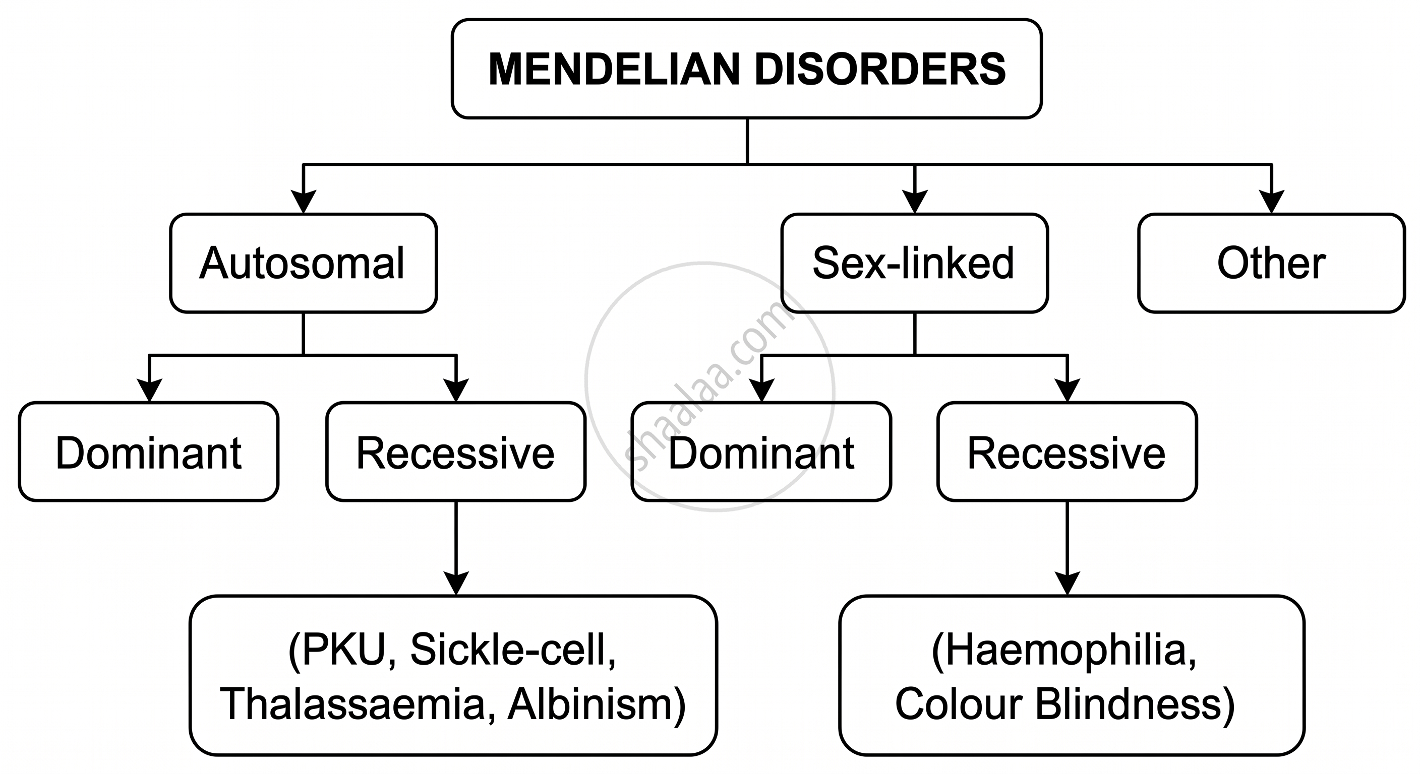
Mendelian disorders are genetic disorders caused by mutations or alterations in a single gene (monogenic disorders). They follow the classical Mendelian patterns of inheritance - autosomal dominant, autosomal recessive, or sex-linked - and can be traced through family pedigrees.
Key Characteristics:
- Caused by alteration/mutation in one gene locus
- Can be identified by pedigree analysis of family history
- Either inherited from one or both parents or arises due to new mutations
- Relatively rare - may affect one in every thousand or a million individuals
CISCE: Class 12
Classification of Mendelian Disorders
CISCE: Class 12
Autosomal Recessive Disorders: Sickle-Cell Anaemia
An autosomal recessive blood disorder caused by a point mutation in the β-globin gene, leading to structurally abnormal haemoglobin (HbS) and sickle-shaped red blood cells.
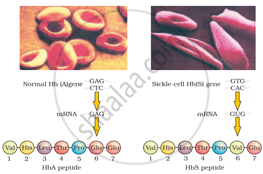
Micrograph of the red blood cells and the amino acid composition of the relevant portion of the β-chain of haemoglobin: (a) From a normal individual; (b) From an individual with sickle-cell anaemia
Gene & Chromosome:
- Gene: HBB (β-globin gene) on Chromosome 11 (short arm)
- Alleles: HbA (normal) and HbS (mutant)
Molecular Mechanism:
| Level | Normal | Mutant (Sickle-cell) |
|---|---|---|
| DNA (codon) | GAG | GTG (or GUG at mRNA level) |
| mRNA codon | GAG | GUG |
| Amino acid (Position 6, β-chain) | Glutamic acid (polar, hydrophilic) | Valine (non-polar, hydrophobic) |
| Haemoglobin | HbA (normal) | HbS (defective) |
Genotypes & Phenotypes:
| Genotype | Condition | Phenotype |
|---|---|---|
| HbA HbA | Normal | Normal RBCs |
| HbA HbS | Carrier (Sickle-cell Trait) | Mostly normal, mild anaemia under stress |
| HbS HbS | Affected (Sickle-cell Disease) | Severe anaemia, sickle-shaped RBCs |
Pathophysiology:
- Under low oxygen conditions, HbS molecules polymerise due to hydrophobic valine forming tactoids (crystalline structures)
- Tactoids distort RBCs into crescent/sickle shapes
- Sickled RBCs adhere to blood vessel walls → vaso-occlusion → tissue damage
- Sickled RBCs are fragile → rupture → haemolytic anaemia
CISCE: Class 12
Autosomal Recessive Disorders: Phenylketonuria (PKU)
An autosomal recessive inborn error of metabolism caused by a deficiency of the enzyme phenylalanine hydroxylase (PAH), leading to the accumulation of phenylalanine and its toxic derivatives.
Gene & Chromosome:
- Gene: PAH on Chromosome 12q (long arm)
- PAH enzyme deficiency accounts for ~95% of PKU cases
Mechanism:
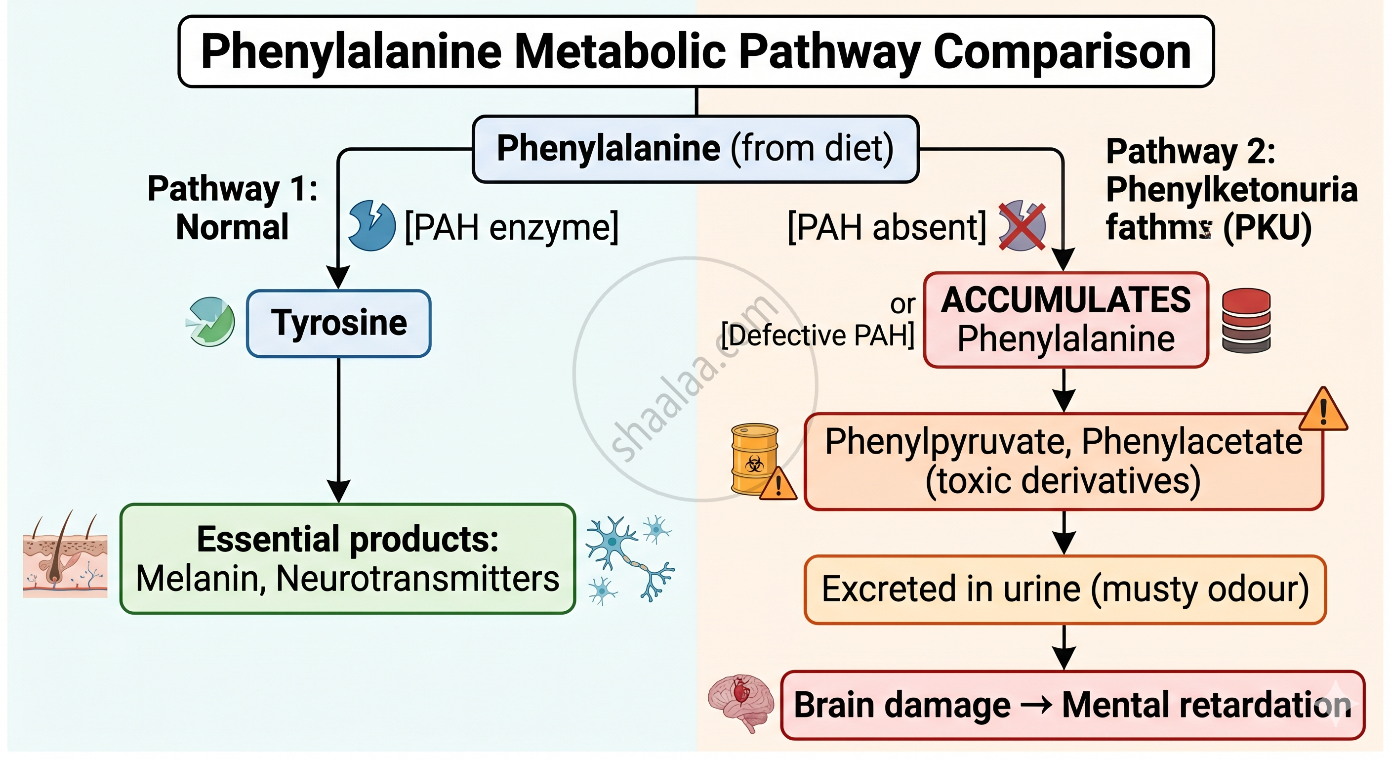
Key Features:
- Mental retardation, if untreated
- Reduced hair and skin pigmentation (less tyrosine → less melanin)
- Phenylpyruvate and phenylacetate are excreted in urine
- Ferric chloride test → green discolouration of urine (diagnostic)
- Newborn screening (Guthrie test / Tandem Mass Spectrometry within 24–48 hours of birth) allows early dietary intervention
CISCE: Class 12
Autosomal Recessive Disorders: Thalassaemia
An autosomal recessive blood disorder characterised by reduced synthesis (quantitative defect) of α- or β-globin chains of haemoglobin, resulting in severe anaemia.
Gene & Chromosome:
- α-Thalassaemia → mutations in HBA1 and HBA2 genes on Chromosome 16
- β-Thalassaemia → mutations in HBB gene on Chromosome 11
Types Comparison:
| Type | Affected Chain | Gene Locus | Key Feature |
|---|---|---|---|
| α-Thalassaemia | α-globin chain | HBA1, HBA2 (Chr 16) | Deletions are common; severity varies by number of deleted genes |
| β-Thalassaemia | β-globin chain | HBB (Chr 11) | Point mutations; β-Thalassaemia Major (Cooley's anaemia) is most severe |
Symptoms: Severe anaemia, bone deformities, facial deformities, enlarged spleen/liver, jaundice, poor growth.
Treatment: Regular blood transfusions, bone marrow transplant in severe cases.
CISCE: Class 12
Autosomal Recessive Disorders: Albinism
An autosomal recessive disorder characterised by the absence of melanin in skin, hair, and eyes due to a non-functional or absent tyrosinase enzyme.
Gene & Chromosome:
- TYR gene on Chromosome 11q14 (tyrosinase gene) for OCA type 1
- Inheritance: Autosomal recessive - the sex of individual does NOT affect occurrence
Mechanism:
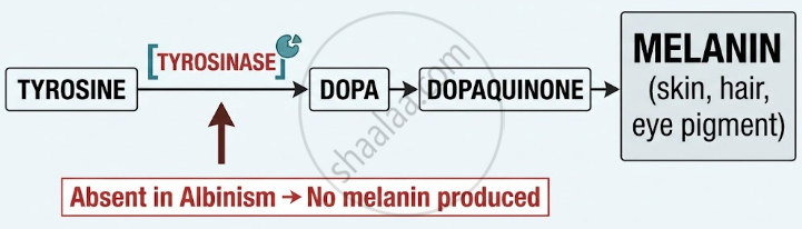
Key Features:
- Very pale/white skin, hair, and eyes
- Photosensitivity - high risk of sunburn and skin cancer
- Vision problems - lack of melanin in the retina
- Carriers (Aa) are normal in appearance; only aa individuals are affected
- Real-Life Note: Approximately 1 in 20,000 people globally are affected by oculocutaneous albinism (OCA)
CISCE: Class 12
X-Linked Recessive Disorders: Haemophilia
An X-linked recessive disorder in which blood clotting is impaired due to a deficiency of clotting factors (Factor VIII in Haemophilia A; Factor IX in Haemophilia B).
Gene & Chromosome:
- Gene located on the X chromosome (sex chromosome)
- Males (XY): Only one X → if defective, disease manifests (hemizygous)
- Females (XX): Need two defective copies to be affected; one copy = carrier
Genotype Table:
| Genotype | Individual | Status |
|---|---|---|
| XH XH | Female | Normal |
| XH Xh | Female | Carrier (normal phenotype) |
| Xh Xh | Female | Affected (very rare) |
| XH Y | Male | Normal |
| Xh Y | Male | Affected (haemophilic) |
Key Features:
- Prolonged/uncontrolled bleeding even from minor cuts
- Internal bleeding in joints (haemarthrosis) is common
- Transmitted from carrier mother to son (criss-cross inheritance)
- Haemophilic father → all daughters are carriers; all sons are normal (if mother is normal)
CISCE: Class 12
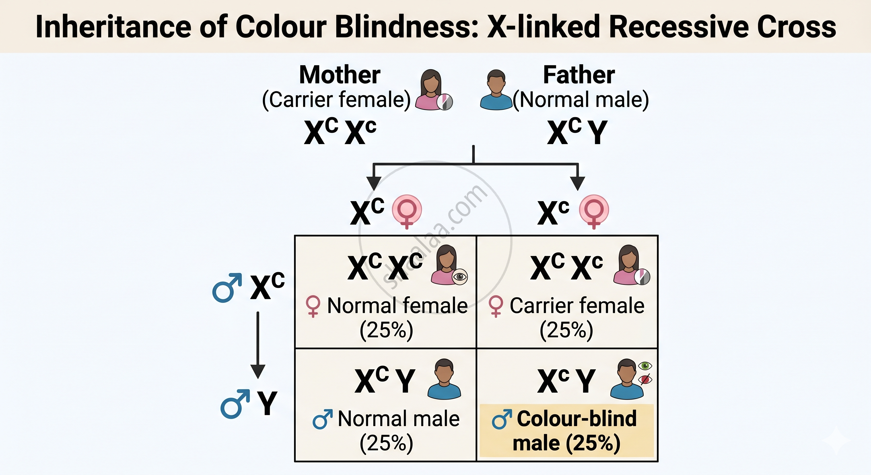
X-Linked Recessive Disorders: Colour Blindness
An X-linked recessive disorder in which the individual cannot distinguish between red and green colours, due to defective or absent cone pigments in the retina.
Gene & Chromosome:
- Genes for red-sensitive (OPN1LW) and green-sensitive (OPN1MW) cone pigments on the X chromosome
- Dominant allele Xc → normal vision; Recessive allele Xc → colour blind
Key Features:
- Affected individuals see red and green as shades of grey
- Approximately 8% of males and 0.4% of females are colour blind (higher in males because males are hemizygous)
- Shows criss-cross inheritance: colour-blind father passes the trait to all daughters (as carriers), not to sons
Cross: Carrier Female × Normal Male
CISCE: Class 12
Key Points: Mendelian Disorders in Humans
| Disorder | Inheritance Type | Chromosome Involved | Main Defect | Key Features |
|---|---|---|---|---|
| Haemophilia | X-linked recessive | X-chromosome | Defective blood-clotting protein | Excessive bleeding from minor cuts; mainly affects males |
| Colour Blindness | X-linked recessive | X-chromosome | Defect in red/green cone pigments | Inability to distinguish red and green colours |
| Sickle-Cell Anaemia | Autosomal recessive | Autosome (Chr 11) | Valine replaces glutamic acid in β-globin | Sickle-shaped RBCs, anaemia, reduced oxygen transport |
| Phenylketonuria (PKU) | Autosomal recessive | Autosome | Lack of enzyme that converts phenylalanine to tyrosine | Mental retardation due to phenylalanine accumulation |
| Thalassaemia | Autosomal recessive | Autosomes (Chr 11 / 16) | Reduced synthesis of α or β globin chains | Severe anaemia, fragile RBCs |
| Albinism | Autosomal recessive | Autosome | Absence of tyrosinase enzyme → no melanin | Very pale skin, hair and eyes; sun sensitivity |
