Advertisements
Advertisements
Question
The figures (A) and (B) given below are showing some kind of adjustment. Study the figures and answer the questions that follow.
| A | 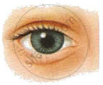 |
 |
B |
- Identify the kinds of adjustments done in the figures (A) and (B).
- Distinguish between the adjustments of figures (A) and (B) on the basis of:
- The size of pupil.
- The pigment which gets regenerated.
- Cells of the retina.
Advertisements
Solution
- The kind of adjustments done in Figure (A) is dilated pupil due to dim light.
The kind of adjustments done in Figure (B) is constricted pupil due to bright light. -
- In the case of A, the pupil dilates to allow more light into the eyes.
In case B, the pupil constricts, reducing the amount of light entering the eyes. - Case A involves the regeneration of visual purple or rhodopsin.
Case B involves the regeneration of cone cells or iodopsin. - Rod cells become activated and begin producing rhodopsin.
Cone cells become activated and begin producing iodopsin.
- In the case of A, the pupil dilates to allow more light into the eyes.
APPEARS IN
RELATED QUESTIONS
The least distance of distinct vision for a young adult with normal vision is about ______.
Why is a normal eye not able to see clearly the objects placed closer than 25 cm?
Write an Explanation.
Persistence of vision
Identify and explain concepts given in this diagram.
The ability of the eye lens to adjust its focal length to form a sharp image of the object at varying distances on the retina is called:
The focal length of the eye lens increases when eye muscles ____________.
How are we able to see nearby and also the distant objects clearly?
In the blind spot of the human eye ______.
Adjusting the focal length of the eye lens to view objects at different distances is done by ______.
The vitamin required for the synthesis of rhodopsin is ______.
