Advertisements
Advertisements
Question
The diagram given below represents the spinal cord of a mammal, seen in a transverse section together with the nerves. Study the diagram and then answer the questions given below:
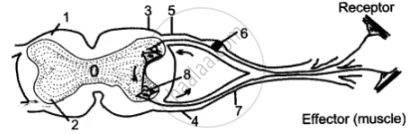
(i) Label the parts 1—8 indicated by guidelines.
(ii) What do the arrows indicate? What is the pathway indicated termed?
(iii) What type of nerve is shown in the diagram?
Advertisements
Solution
(i)
- White matter
- Gray matter
- Dorsal root
- Ventral root
- Dorsal ganglion
- Sensory neuron
- Spinal nerve
- Synapse
(ii) The arrows indicate the arrangement of white and gray matter is reversed from that in the brain. The patl4way indicated is termed as a nerve pathway in a simple spinal reflex.
(iii) Sensory or Afferent nerve.
APPEARS IN
RELATED QUESTIONS
Distinguish between the following pair of words:
Cranial nerve and spinal nerve
Where is this located?
Meninges
Where is this located?
Ganglia
Give one point of difference between the following on the basis of what is indicated:
Cerebrum and spinal cord. (Arrangement of neurons)
Spinal cord and sympathetic ganglion of autonomous nervous system are connected by ______________.
The diagram given below shows the internal structure of the spinal cord, depicting a simple reflex. Study the same and then answer the questions that follow:
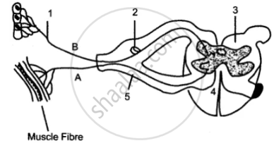
(i) Name the parts numbered 1 to 5.
(ii) Using the letters of the alphabet shown in the figure indicate the direction in which an impulse enters and leaves the spinal cord.
(iii) What is the term given to the point of contact between two nerve cells?
(iv) What is meant by ‘simple reflex’ ? Give two examples of simple reflexes and name the stimuli too.
(v) How does the arrangement of nerve cells in the spinal cord differ from that in the brain?
Identify the odd item from the following series of words.
Sketch and label T. S. of Spinal cord.
There are ______ pairs of cranial nerves and ______ pairs of spinal nerves.
In humans, the number of spinal nerves are ______.
