Advertisements
Advertisements
प्रश्न
The diagram given below represents the spinal cord of a mammal, seen in a transverse section together with the nerves. Study the diagram and then answer the questions given below:
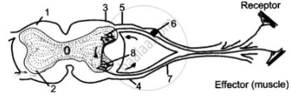
(i) Label the parts 1—8 indicated by guidelines.
(ii) What do the arrows indicate? What is the pathway indicated termed?
(iii) What type of nerve is shown in the diagram?
Advertisements
उत्तर
(i)
- White matter
- Gray matter
- Dorsal root
- Ventral root
- Dorsal ganglion
- Sensory neuron
- Spinal nerve
- Synapse
(ii) The arrows indicate the arrangement of white and gray matter is reversed from that in the brain. The patl4way indicated is termed as a nerve pathway in a simple spinal reflex.
(iii) Sensory or Afferent nerve.
APPEARS IN
संबंधित प्रश्न
Name the following:
Inability to speak.
Where is this located?
Ganglia
Complete the following sentence with appropriate word :
The part of the central nervous system which responds to the reflex action is ________.
Mention, if the following statement is True or False
Spinal nerves are twelve pair
Sketch and label T. S. of Spinal cord.
There are ______ pairs of cranial nerves and ______ pairs of spinal nerves.
Describe the structure of the spinal cord.
Spinal cord is enclosed in ____________ of vertebral column.
Spinal nerves are usually ______
Mention where in the human body is the following located and state its main function:
Central canal
