Advertisements
Advertisements
Question
The diagram given below shows the internal structure of the spinal cord, depicting a simple reflex. Study the same and then answer the questions that follow:
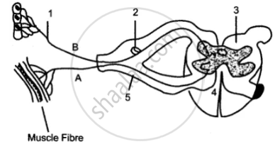
(i) Name the parts numbered 1 to 5.
(ii) Using the letters of the alphabet shown in the figure indicate the direction in which an impulse enters and leaves the spinal cord.
(iii) What is the term given to the point of contact between two nerve cells?
(iv) What is meant by ‘simple reflex’ ? Give two examples of simple reflexes and name the stimuli too.
(v) How does the arrangement of nerve cells in the spinal cord differ from that in the brain?
Advertisements
Solution
(i)
1. Sensory or Afferent fiber.
2. The cell body of the sensory neurons.
3. White matter.
4. The cell body of the motor neurons.
5. Spinal nerve.
(ii) BCA
(iii) Synapse.
(iv) Simple reflex: It is an involuntary automatic, quick response to a stimulus.
Examples:
(a) The closing of the eyelids when a strong beam of light is flashed.
(b) The withdrawal of the hand when it is pricked.
(v) In spinal cord white matter is outside and gray matter is inside. In brain white matter is inside and gray matter is outside.
APPEARS IN
RELATED QUESTIONS
Distinguish between the following pair of words:
Motor and sensory nerve
Choose the correct answer.
Number of spinal nerves in human beings ______________
The diagram given below represents the spinal cord of a mammal, seen in a transverse section together with the nerves. Study the diagram and then answer the questions given below:
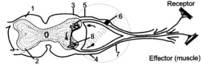
(i) Label the parts 1—8 indicated by guidelines.
(ii) What do the arrows indicate? What is the pathway indicated termed?
(iii) What type of nerve is shown in the diagram?
Complete the following sentence with appropriate word :
The part of the central nervous system which responds to the reflex action is ________.
Identify the odd item from the following series of words.
There are ______ pairs of cranial nerves and ______ pairs of spinal nerves.
Sensory nerve fibres forming ascending tracts in white matter of spinal cord are located in ____________.
______ connects the brain to a different part of the body through nerves.
How are cytons and axons arranged in the spinal cord?
Explain the formation of a typical spinal nerve with the help of a neat labelled diagram.
