Advertisements
Advertisements
Question
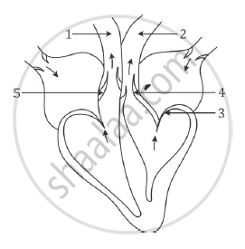
The diagram below represents the human heart in one phase of its function. Study the diagram carefully and answer the questions that follow:
(i) Name the phase.
(ii) Which part of the heart is contracting in this phase? Give a reason to support your answer.
(iii) Name the parts labelled 1 to 4.
(iv) What type of blood flows through ‘2’?
(v) State the function of the part numbered ‘5’.
(vi) Name the membrane that covers the heart.
Advertisements
Solution
(i) Ventricular systole
(ii) Ventricles are contracting in this phase. In the diagram given, tricuspid valves and bicuspid valves are closed, while the semi-lunar valves are open.
(iii)
1 – Pulmonary artery
2 – Aorta
3 – Bicuspid valve
4 – Semilunar valve (aortic semilunar valve)
(iv) Oxygenated blood flows through ‘2’, i.e. aorta.
(v) ‘5’ is pulmonary semilunar valve. It prevents the backflow of blood into the right ventricle at the time of ventricular diastole.
(vi) Pericardium covers the heart.
APPEARS IN
RELATED QUESTIONS
Aqueous humour, Vitreous humour, lris, Central canal
What are the average values of blood pressure in a normal adult human?
Give reason for the following:
The blood groups of both the donor and recipient must be known before transfusing blood.
pH of oxygenated blood is ______.
Put a tick mark (✓) against the correct alternative in the following statement.
Pulmonary vein carries
Fill in the blank:
______ carry pure blood.
True or false:
People with blood group ‘O’ are called universal recipients
Complete the following sentence with appropriate word:
The membranous covering of the heart is _______.
The heart is covered by ______.
Given diagram is a schematic representation of the circulatory system in humans. Study the same and answer the questions that follow:
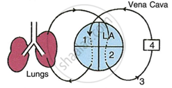 |
- Label the parts 1 and 4 indicated in the diagram.
- Which of the above mentioned number is the thickest artery? Also write its name.
- Mention the number and chamber of the heart which has the thickest muscular wall.
- Which of the above numbers/structures has the maximum number of blood capillaries?
- Draw neat and labelled diagrams of the transverse section of vena cava and the part numbered as 3. Make sure to show the structural differences between these two in the diagram.
