Advertisements
Advertisements
Question
Match structures given in Column I with functions given in Column II.
| Column I | Column II | ||
| (i) | Stomata | (a) | Absorption of water |
| (ii) | Xylem | (b) | Transpiration |
| (iii) | Root hairs | (c) | Transport of food |
| (iv) | Phloem | (d) | Transport of water |
| (e) | Synthesis of carbohydrates |
Advertisements
Solution
| Column I | Column II | ||
| (i) | Stomata | (b) | Transpiration |
| (ii) | Xylem | (d) | Transport of water |
| (iii) | Root hairs | (a) | Absorption of water |
| (iv) | Phloem | (c) | Transport of food |
APPEARS IN
RELATED QUESTIONS
Bile, Urea, Uric acid, Ammonia
_____________ secreted by the (ii) _____________ lobe of the pituitary gland. If this hormone secretion is reduced, there is an increased production of urine. This disorder is called (iii) ____________. Sometimes excess glucose is passed with urine due to hyposecretion of another hormone called (iv) _____________ leading to the cause of a disease called (v) ______________.
What gaseous waste products are excreted by plants?
With which human organ system (or human systems) is the glomerulus associated?
Write down the functional activity of the following parts,
Henle's loop ………………
Choose the correct answer:
Composition of extracellular fluid is regulated by ___________
Choose the correct option.
The part of nephron which absorbs glucose and amino acid is ______.
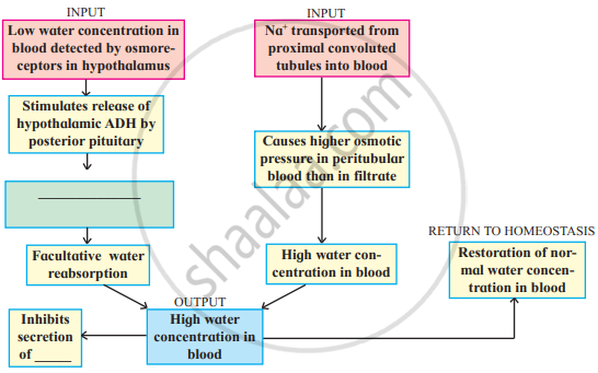
Complete the diagram/chart with correct labels/ information. Write the conceptual details regarding it.
The CORRECT match is ______.
Name the organs that store and release the urine.
