Advertisements
Advertisements
Question
Explain the structure of secondary oocyte.
Advertisements
Solution

The secondary oocyte is the unfertilized egg which is released from the ovary (ovulated).
- Size and structure:
It is non-cleidoic (without shell) and microlecithal (yolk is present in very small quantities). It is approximately 0.1mm (100 microns) in size. It is a rounded, nonmotile, haploid female gamete. The egg shows polarity i.e. the side having a germinal vesicle and the first polar body is called an animal pole while the side opposite to it is called the vegetal pole. - Nucleus:
The nucleus of the egg appears large and is called a germinal vesicle. A typical nucleus or pronucleus is formed at the time of fertilization. - Cytoplasm:
The cytoplasm of the egg is also called ooplasm. It is devoid of centrioles. - Membranes:
The egg is surrounded by various coverings. The egg membrane is called the vitelline membrane. It secretes a non-cellular, glycoproteinous membrane with the zona pellucida on its outside. Adhering to the outer surface of zona pellucida are several radially elongated cells forming the corona radiata. These cells are derived from the innermost layer of granulosa cells. They are firmly held to the zona pellucida and to each other by hyaluronic acid (mucopolysaccharide). Between the vitelline membrane and the zona pellucida is a fluid-filled perivitelline space. The first polar body lies in this space.
APPEARS IN
RELATED QUESTIONS
Body breaks up into several fragments and each fragment starts to live as a new individual. This is ______ type of reproduction.
Multiple choice question.
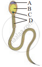
The given figure shows human sperm. Various parts of it are labelled as A, B, C, and D. Which labelled part represents acrosome?
Describe the process of spermatogenesis with the help of diagram.
The male sex hormone testosterone is secreted from ____________.
Mammalian egg is ____________.
It The cell division in secondary oocyte is suspended at ______.
During embryonic development, the establishment of polarity along anterior/posterior, dorsal/ventral or medial/lateral axis is called ______.
Preparation of sperm before penetration of ovum is ______.
During spermatogenesis meiosis occurs in ______.
The diagram given below shows the various steps in spermatogenesis.

- Name the parts labelled '1', '2' and '3'.
- Name the process by which part '3' changes to part '4'.
