Advertisements
Advertisements
Question
Describe the structure of a seminiferous tubule.
Advertisements
Solution
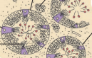
The production of sperm in the testes takes place in a highly coiled structure called the seminiferous tubules. These tubules are located in the testicular lobules. Each seminiferous tubule is lined by germinal epithelium. It is lined on its inner surface by two types of cells: spermatogonia and Sertoli cells. Spermatogonia are male germ cells that give rise to primary spermatocytes through meiotic divisions. Primary spermatocytes undergo further meiotic division to form secondary spermatocytes and finally, spermatids. Spermatids later metamorphose into male gametes called spermatozoa. Sertoli cells are known as nurse cells of the testes, as they provide nourishment to the germ cells. There are large polygonal cells known as interstitial cells or Leydig cells just adjacent to the seminiferous tubules. These cells secrete the male hormone called testosterone.

APPEARS IN
RELATED QUESTIONS
Write the location and functions of the following in human testes :
Leydig cells
Identify True/False statement. Correct the false statement to make it true.
Leydig cells are found in the ovary.
find odd one from following: Thymine, Cytosine, Adenine, Pepsin.
List the structures, in their correct sequence, through which the sperms must pass from the time they are produced in the testes to the time they leave the urethra.
Define the following term:
Hernia
With the help of labelled diagramms describe the structures of human sperm and unfertilized ovum.
A couple is unable to conceive. Which modern techniques are available to overcome this problem?
What differences are there in number, structure, and activity of the male and female gametes in a man?
Explain the Term Penis.
Explain the term Scrotum.
Name the Following
The hormone that stimulates the development of secondary sexual characters in males.
Write the functional activity of the following structure:
Cowper’s gland
The increased level of estrogen and progesterone is responsible for menstruation.
The given diagram represents T. S of
In the following diagram, what are A and its function?

Select the MISMATCHED pair.
Failure of testes to descend from abdomen into scrotum leads to ______.
Which of the following is a transporting tube leading from the bladder to which brings urine outside the body via penis?
In humans, male germs cells differentiate into ______ at the end of first meiotic division.
In human males, the testes lie in the scrotum, because it helps in the ______
In the following Question, the Assertion and Reason have been put forward. Read the statements carefully and choose the correct alternative from the following:
Assertion: In the male reproductive system, the transport of sperm takes place in a fluid that also provides nutrition.
Reason: Protective glands and seminal vesicles secret in the vas deferens.
Crypts of Lieberkuhn are absent in ______.
The process of formation of sperms is known as ______.
What are the components of semen?
What is the significance of the testes being located in scrotal sacs outside the abdomen?
Mention the location and the function of Leydig cells.
Answer the following question based on the human male reproductive system:
What is the work of vas deferens?
Answer the following question based on the human male reproductive system:
By which type of cell division, sperms are formed?
The male accessory gland whose secretion neutralizes the acidity of the urethra and vagina is ______.
The canal through which each testis descends into the scrotum just before the birth of a male baby is ______.
The figure given below is an important gonad of humans. Study the figure and answer the following questions.
 |
- Identify the organ. Write its specific location in the body.
- Label the parts shown in the figure as 1 to 4.
- Write important functions of parts 2 and 4.
- Name one cellular structure and one hormone which are produced in part 3.
- Draw a neat and labelled diagram of the cellular structure mentioned by you in (d).
