Advertisements
Advertisements
प्रश्न
Write briefly about the Testis.
Advertisements
उत्तर
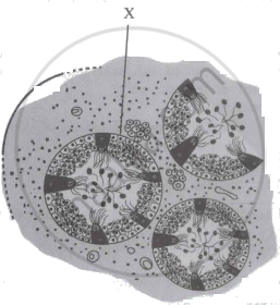
The testis is externally covered by a collagenous connective tissue layer called the tunica albuginea. Outer to it is an incomplete peritoneal covering called tunica vaginalis, and inner to it is tunica vasculosa, a thin membranous and vascular layer. Fibers from tunica albuginea divide each testis into about 200-300 testicular lobules. Each with 1-4 highly coiled seminiferous tubules. Each seminiferous tubule is internally lined by cuboidal germinal epithelial cells (spermatogonia) and few large pyramidal cells called Sertoli or sustentacular cells. The germinal epithelial cells undergo gametogenesis to form spermatozoa. Sertoli cells provide nutrition to the developing sperms. Various stages of spermatogenesis can be seen in the seminiferous tubules. The inner most spermatogonial cell (2n), primary spermatocyte (2n), secondary spermatocyte (n), spermatids (n) and sperms (n). The Interstitial or Leydig’s cells lie in between the seminiferous tubules. They secrete the male hormone androgen or testosterone.
APPEARS IN
संबंधित प्रश्न
Differentiate between Rod cells and cone cells (pigment)
Write the functional activity of the following structure:
Seminiferous tubules
The increased level of estrogen and progesterone is responsible for menstruation.
Identify the primary sex organs in males.
In the given diagram represents the
Vasa efferentia are the ductules leading from ______.
Secretions from which one of the following are rich in fructose, calcium, and some enzymes?
Seminal plasma, the fluid part of semen is formed by ______.
A pair of ducts arising from testis, which carries sperms are ______
The loose fold of skin covering the glans penis is called the:
