Advertisements
Advertisements
प्रश्न
Explain the formation of a typical spinal nerve with the help of a neat labelled diagram.
Advertisements
उत्तर

Formation of Spinal Nerve
- All spinal nerves are of the mixed type i.e. they have some nerve fibre as sensory and some motor.
- Each spinal nerve is formed inside the neural canal of vertebral column by two roots - the posterior or dorsal sensory root and anterior or ventral root.
- Anterior root receives the sensory nerve from the dorsal root ganglion (cell bodies of sensory neurons are located in the ganglion), while the anterior/ventral root gives out the motor nerve.
- The dorsal sensory and the ventral motor nerves together form the mixed spinal nerve.
- It emerges out from both sides of the spinal cord through the intervertebral foramen.
- As soon as it emerges out of vertebral column, it shows three branches viz.
- Ramus dorsalis: From skin and to muscles of dorsal side.
- Ramus ventralis: The largest of the three supplies the organs and muscles on lateral and anterior side.
- Ramus communicans: The smallest of the three and given out from 1st thoracic upto 3rd lumbar (L3) spinal nerve. It joins the sympathetic ganglia.
APPEARS IN
संबंधित प्रश्न
Differentiate between following pair with reference to the aspect in bracket.
cerebrum and spinal cord (arrangement of cytons and exons of neurons).
Name the following:
Inability to speak.
Choose the correct answer.
Number of spinal nerves in human beings ______________
Give Technical Term:
The fluid which fills the central canal of the spinal cord.
The diagram given below shows the internal structure of the spinal cord, depicting a simple reflex. Study the same and then answer the questions that follow:
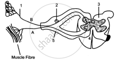
(i) Name the parts numbered 1 to 5.
(ii) Using the letters of the alphabet shown in the figure indicate the direction in which an impulse enters and leaves the spinal cord.
(iii) What is the term given to the point of contact between two nerve cells?
(iv) What is meant by ‘simple reflex’ ? Give two examples of simple reflexes and name the stimuli too.
(v) How does the arrangement of nerve cells in the spinal cord differ from that in the brain?
Describe the structure of the spinal cord.
Sensory nerve fibres forming ascending tracts in white matter of spinal cord are located in ____________.
Spinal cord is enclosed in ____________ of vertebral column.
How are cytons and axons arranged in the spinal cord?
Spinal nerves are usually ______
