Advertisements
Advertisements
प्रश्न
The help of a well-labelled diagram describes the internal structure of the human heart.
Advertisements
उत्तर

Human heart has four chambers, two atria and two ventricles.
Atria:
Right atrium:
- Two atria are separated by a septum. The right atrium receives deoxygenated blood from upper part of the body by superior vena cava and inferior vena cava collects blood from he lower part of the body. Coronary sinus brings blood from the heart muscles.
- Eustachian valve guards the opening of inferior vena cava while the besian valve is present near the opening of coronary sinus.
- A dperession called fossa ovails is present on the right side of interatrial septum.
- Right atrium opens into right ventricle.
Left atrium:
- Oxygeneated blood from lungs comes here via pulmonary veins.
- Left atrium opens into left ventricle.
Ventricles:
- Ventricles are the distributing chambers which are separated by interventricular septum.
- Left ventricle has thickest wall as it pumps blood to all parts of the body.
- In the opening of right atrium into right ventricle, tricuspid valve (three flaps) is present which controls the transportation of blood from right atrium to right ventricle. Similarly bicuspid valve is present in between left atrium and left venticle. It is also known as mitral valve.
- These valves are attached to the ventricle by chorac tendineae, which prevents the back flow of blood.
From right ventricle: Pulmonary trunk carries deoxygenated blood to lungs for oxygenation.
From left ventricle: The aorta distributes oxygenated blood to all parts of the body. Pulmonary aorta and systematic aorta have three semilunar valves at the base which prevents back flow of blood.
APPEARS IN
संबंधित प्रश्न
Fill in the blank.
In human body, heart is located on the _____ side of the chest cavity.
Why is the SA node called the pacemaker of the heart?
Draw the diagram of the Position of valves in the human heart.
Explain the Term
Pace maker
State the Function: Semilunar valves of the heart
In between which layers of pericardium, pericardial fluid is present?
Draw diagram of conducting system of human heart. Label SA node and bundle of His.
What is the importance of valves in the heart?
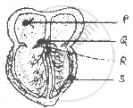
Observe the diagram, Identify the parts labelled as P, Q, R, S. Choose the correct option for the conduction of cardiac impulse.
Assertion: Human heart does not allow the mixing of oxygen to reach blood with carbon dioxide to reach the blood.
Reason: Human heart has different chambers.
