Advertisements
Advertisements
प्रश्न
Given below is a diagram of the external features of the human heart. Study the figure and answer the questions that follows:
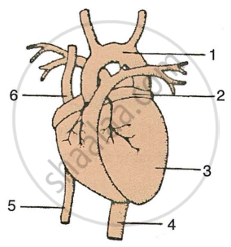 |
- Label the guidelines shown as 1 to 6 in the figure.
- Write the important role of parts 5 and 6.
- Name the chamber of the heart which collects blood from the lungs through a blood vessel. Also write the name of the blood vessel.
- Write one structural and one functional difference between the blood vessels 4 and 5.
- What happens when there is a blockage in any coronary artery or any of their branches?
दीर्घउत्तर
Advertisements
उत्तर
-
- Aorta
- Left Atrium
- Left Ventricle
- Dorsal Aorta
- Inferior Vena Cava
- Superior Vena Cava
- The vena cava is a vein that transports deoxygenated blood from the body to the heart. There are two vena cava, superior and inferior. The superior vena cava transports deoxygenated blood from the upper body, including the head, arms, and neck. In contrast, the inferior vena cava transports it from the lower body, including the legs and abdominal organs.
- The left atrium receives blood from the lungs through pulmonary veins.
- The structural difference between the inferior vena cava and the dorsal aorta.
The inferior vena cava has valves and a broader lumen, while the dorsal aorta has no valves and a limited canal.
The functional distinction between the inferior Vena Cava and the dorsal aorta.
The dorsal aorta transports oxygenated blood from the heart's left ventricle to the body, while the vena cava drains deoxygenated blood to the right ventricle. - A coronary artery blockage or branch can restrict blood flow to the heart muscle. Reduced blood flow can cause chest pain (angina) and, in severe situations, heart attack (myocardial infarction) by damaging the heart muscle. The severity of the repercussions varies depending on the size of the blockage and impacted blood vessels.
shaalaa.com
या प्रश्नात किंवा उत्तरात काही त्रुटी आहे का?
